3FEV image
Entry Detail
PDB ID:
3FEV
Keywords:
Title:
Crystal structure of the chimeric muscarinic toxin MT7 with loop 1 from MT1.
Biological Source:
Source Organism:
PDB Version:
Deposition Date:
2008-12-01
Release Date:
2009-12-22
Method Details:
Experimental Method:
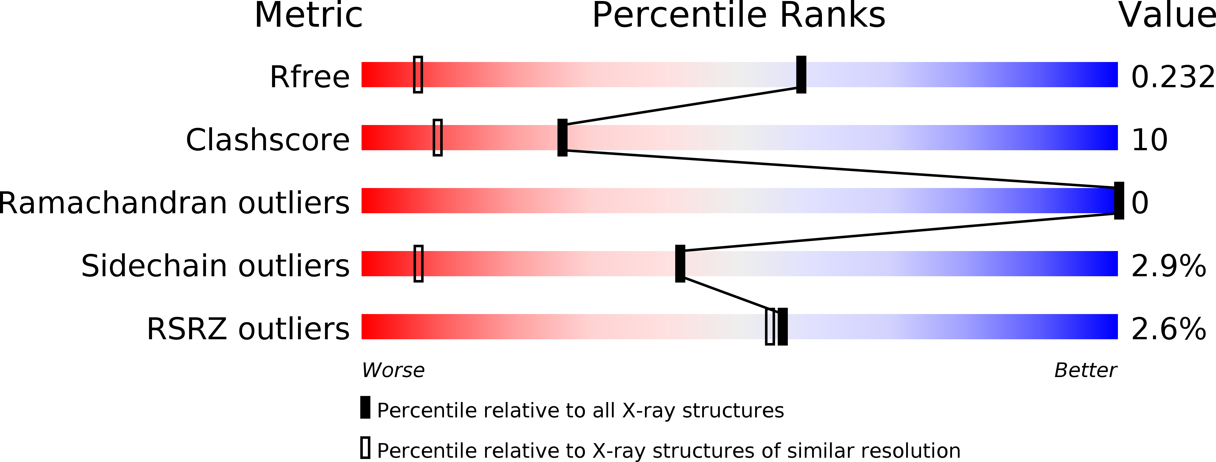
Resolution:
1.30 Å
R-Value Free:
0.23
R-Value Work:
0.21
R-Value Observed:
0.21
Space Group:
P 21 21 21
Macromolecular Entities
Polymer Type:polypeptide(L)
Description:Fusion of Muscarinic toxin 1, Muscarinic m1-toxin1
Chain IDs:A, B, C
Chain Length:65
Number of Molecules:3
Biological Source:Dendroaspis angusticeps
Ligand Molecules
Primary Citation
Engineering of three-finger fold toxins creates ligands with original pharmacological profiles for muscarinic and adrenergic receptors.
Plos One 7 e39166 e39166 (2012)
PMID: 22720062 DOI: 10.1371/journal.pone.0039166

Abstact

Protein engineering approaches are often a combination of rational design and directed evolution using display technologies. Here, we test "loop grafting," a rational design method, on three-finger fold proteins. These small reticulated proteins have exceptional affinity and specificity for their diverse molecular targets, display protease-resistance, and are highly stable and poorly immunogenic. The wealth of structural knowledge makes them good candidates for protein engineering of new functionality. Our goal is to enhance the efficacy of these mini-proteins by modifying their pharmacological properties in order to extend their use in imaging, diagnostics and therapeutic applications. Using the interaction of three-finger fold toxins with muscarinic and adrenergic receptors as a model, chimeric toxins have been engineered by substituting loops on toxin MT7 by those from toxin MT1. The pharmacological impact of these grafts was examined using binding experiments on muscarinic receptors M1 and M4 and on the α(1A)-adrenoceptor. Some of the designed chimeric proteins have impressive gain of function on certain receptor subtypes achieving an original selectivity profile with high affinity for muscarinic receptor M1 and α(1A)-adrenoceptor. Structure-function analysis supported by crystallographic data for MT1 and two chimeras permits a molecular based interpretation of these gains and details the merits of this protein engineering technique. The results obtained shed light on how loop permutation can be used to design new three-finger proteins with original pharmacological profiles.

Legend

Protein

Chemical

Disease

Primary Citation of related structures