Deposition Date
2008-11-21
Release Date
2008-12-16
Last Version Date
2023-12-27
Entry Detail
PDB ID:
3FCG
Keywords:
Title:
Crystal Structure Analysis of the Middle Domain of the Caf1A Usher
Biological Source:
Source Organism(s):
Yersinia pestis (Taxon ID: 632)
Expression System(s):
Method Details:
Experimental Method:
Resolution:
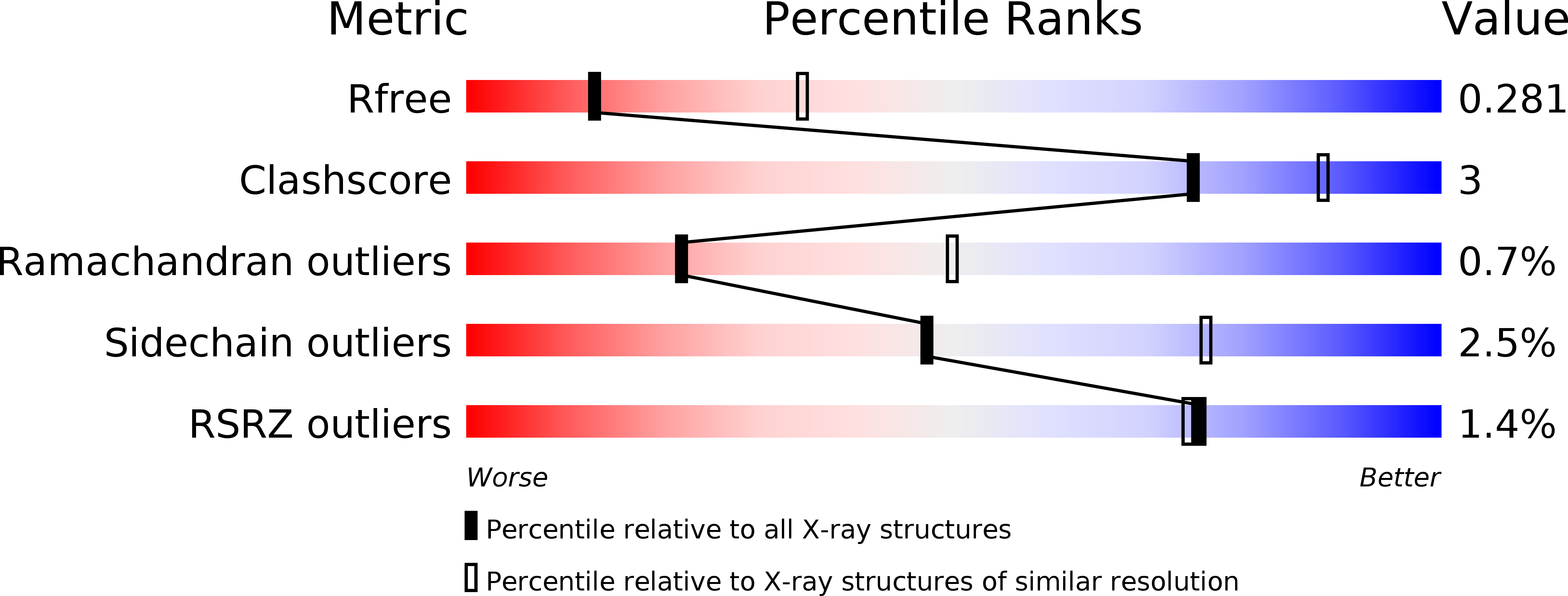
2.85 Å
R-Value Free:
0.28
R-Value Work:
0.26
R-Value Observed:
0.26
Space Group:
C 2 2 21