Deposition Date
2008-11-20
Release Date
2008-12-30
Last Version Date
2024-10-16
Entry Detail
PDB ID:
3FC0
Keywords:
Title:
1.8 A crystal structure of murine GITR ligand dimer expressed in Drosophila melanogaster S2 cells
Biological Source:
Source Organism(s):
Mus musculus (Taxon ID: 10090)
Expression System(s):
Method Details:
Experimental Method:
Resolution:
1.76 Å
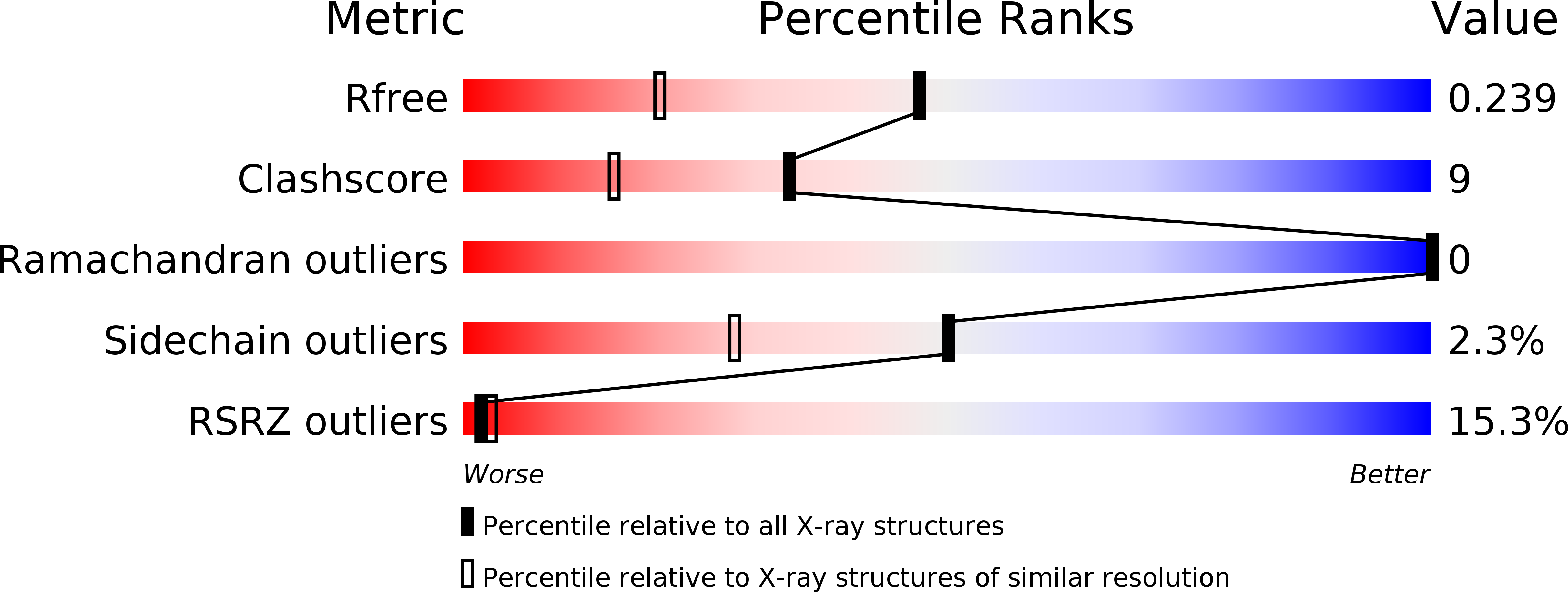
R-Value Free:
0.26
R-Value Work:
0.22
R-Value Observed:
0.22
Space Group:
P 21 21 21