3FB1 image
Deposition Date 2008-11-18
Release Date 2009-11-24
Last Version Date 2023-12-27
Entry Detail
PDB ID:
3FB1
Keywords:
Title:
Crystal Structure of Purine Nucleoside Phosphorylase in Complex with Ribose-1-Phosphate
Biological Source:
Source Organism:
Host Organism:
Method Details:
Experimental Method:
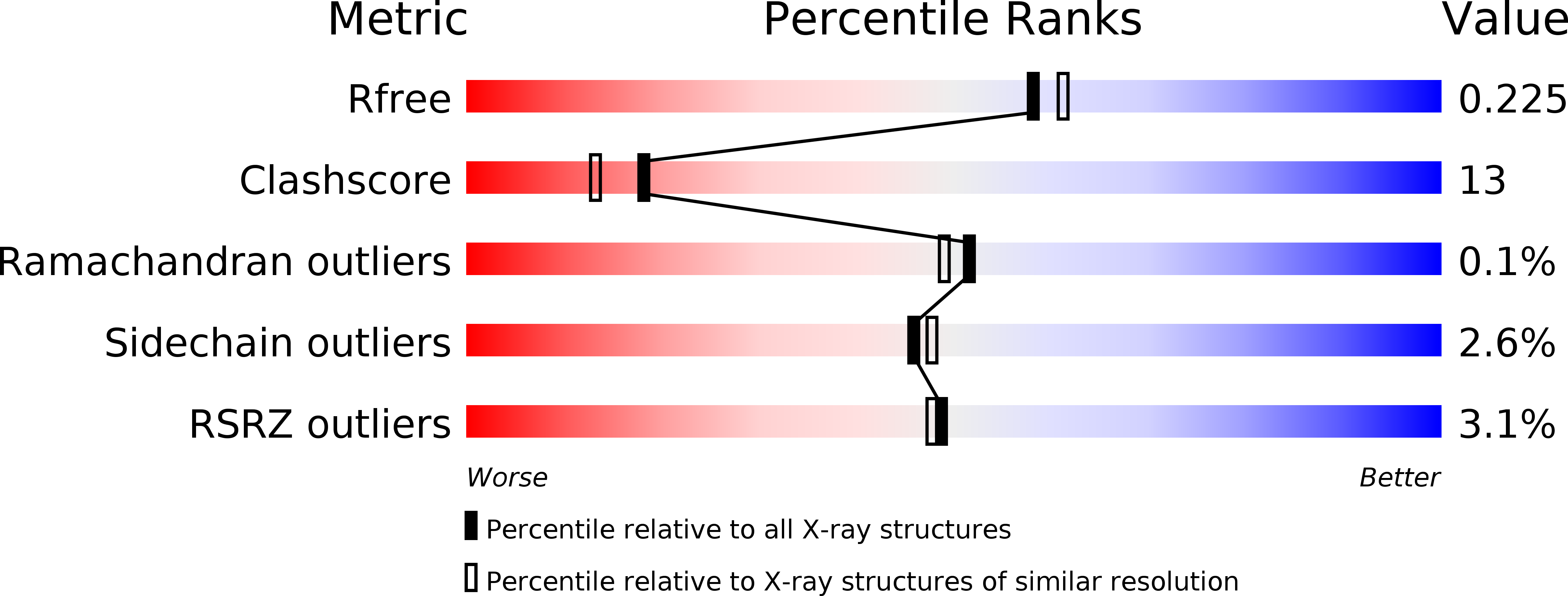
Resolution:
2.00 Å
R-Value Free:
0.23
R-Value Work:
0.17
R-Value Observed:
0.17
Space Group:
P 21 21 21
Macromolecular Entities
Structures with similar UniProt ID
Protein Blast
Polymer Type:polypeptide(L)
Molecule:Purine-nucleoside phosphorylase
Chain IDs:A, B, C
Chain Length:287
Number of Molecules:3
Biological Source:Schistosoma mansoni
Primary Citation
Purine nucleoside phosphorylase from Schistosoma mansoni in complex with ribose-1-phosphate.
J.Synchrotron Radiat. 18 62 65 (2011)
PMID: 21169694 DOI: 10.1107/S0909049510027718

Abstact

Schistosomes are blood flukes which cause schistosomiasis, a disease affecting approximately 200 million people worldwide. Along with several other important human parasites including trypanosomes and Plasmodium, schistosomes lack the de novo pathway for purine synthesis and depend exclusively on the salvage pathway for their purine requirements, making the latter an attractive target for drug development. Part of the pathway involves the conversion of inosine (or guanosine) into hypoxanthine (or guanine) together with ribose-1-phosphate (R1P) or vice versa. This inter-conversion is undertaken by the enzyme purine nucleoside phosphorylase (PNP) which has been used as the basis for the development of novel anti-malarials, conceptually validating this approach. It has been suggested that, during the reverse reaction, R1P binding to the enzyme would occur only as a consequence of conformational changes induced by hypoxanthine, thus making a binary PNP-R1P complex unlikely. Contradictory to this statement, a crystal structure of just such a binary complex involving the Schistosoma mansoni enzyme has been successfully obtained. The ligand shows an intricate hydrogen-bonding network in the phosphate and ribose binding sites and adds a further chapter to our knowledge which could be of value in the future development of selective inhibitors.

Legend

Protein

Chemical

Disease

Primary Citation of related structures