Deposition Date
2008-11-18
Release Date
2008-12-09
Last Version Date
2024-10-16
Entry Detail
PDB ID:
3FAT
Keywords:
Title:
X-ray structure of iGluR4 flip ligand-binding core (S1S2) in complex with (S)-AMPA at 1.90A resolution
Biological Source:
Source Organism(s):
Rattus norvegicus (Taxon ID: 10116)
Expression System(s):
Method Details:
Experimental Method:
Resolution:
1.90 Å
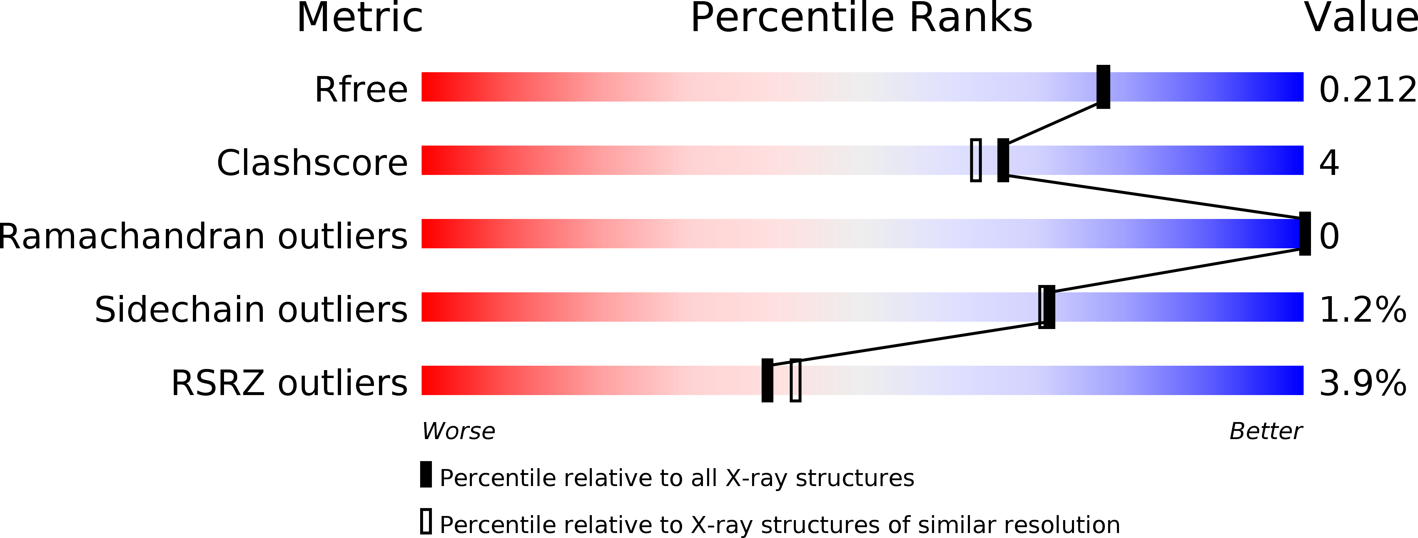
R-Value Free:
0.21
R-Value Work:
0.17
R-Value Observed:
0.17
Space Group:
C 1 2 1