3F95 image
Deposition Date 2008-11-13
Release Date 2009-11-17
Last Version Date 2024-10-16
Entry Detail
PDB ID:
3F95
Keywords:
Title:
Crystal Structure of Extra C-terminal Domain (X) of Exo-1,3/1,4-beta-glucanase (ExoP) from Pseudoalteromonas sp. BB1
Biological Source:
Source Organism:
Host Organism:
Method Details:
Experimental Method:
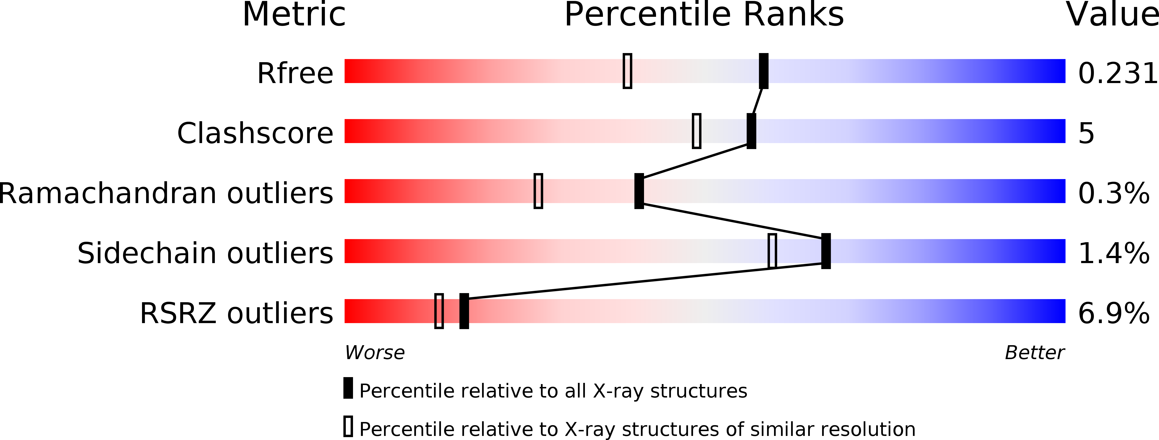
Resolution:
1.80 Å
R-Value Free:
0.23
R-Value Work:
0.18
R-Value Observed:
0.19
Space Group:
P 21 21 2
Macromolecular Entities
Structures with similar UniProt ID
Protein Blast
Polymer Type:polypeptide(L)
Molecule:Beta-glucosidase
Gene (Uniprot):exoP
Chain IDs:A, B
Chain Length:193
Number of Molecules:2
Biological Source:Pseudoalteromonas sp.
Ligand Molecules
Primary Citation
Structure and activity of exo-1,3/1,4-beta-glucanase from marine bacterium Pseudoalteromonas sp. BB1 showing a novel C-terminal domain
Febs J. ? ? ? (2011)
PMID: 22129429 DOI: 10.1111/j.1742-4658.2011.08439.x

Abstact

UNLABELLED Following the discovery of an exo-1,3/1,4-β-glucanase (glycoside hydrolase family 3) from a seaweed-associated bacterium Pseudoalteromonas sp. BB1, the recombinant three-domain protein (ExoP) was crystallized and its structure solved to 2.3 Å resolution. The first two domains of ExoP, both of which contribute to the architecture of the active site, are similar to those of the two-domain barley homologue β-d-glucan exohydrolase (ExoI) with a distinctive Trp-Trp clamp at the +1 subsite, although ExoI displays broader specificity towards β-glycosidic linkages. Notably, excision of the third domain of ExoP results in an inactive enzyme. Domain 3 has a β-sandwich structure and was shown by CD to be more temperature stable than the native enzyme. It makes relatively few contacts to domain 1 and none at all to domain 2. Two of the domain 3 residues involved at the interface, Q683 (forming one hydrogen bond) and Q676 (forming two hydrogen bonds) were mutated to alanine. Variant Q676A retained about half the activity of native ExoP, but the Q683A variant was severely attenuated. The crystal structure of Q683A-ExoP indicated that domain 3 was highly mobile and that Q683 is critical to the stabilization of ExoP by domain 3. Small-angle X-ray scattering data lent support to this proposal. Domain 3 does not appear to be an obvious carbohydrate-binding domain and is related neither in sequence nor structure to the additional domains characterized in other glycoside hydrolase 3 subgroups. Its major role appears to be for protein stability but it may also help orient substrate. DATABASE Structural data are available in the Protein Data Bank under the accession numbers 3UT0, 3USZ, 3F95 and 3RRX.

Legend

Protein

Chemical

Disease

Primary Citation of related structures