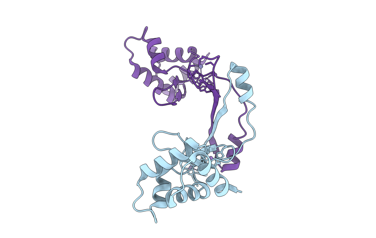
3F8N image
Entry Detail
PDB ID:
3F8N
Title:
Crystal structure of PerR-Zn-Mn
Biological Source:
Source Organism:
Host Organism:
PDB Version:
Deposition Date:
2008-11-13
Release Date:
2009-06-16
Method Details:
Experimental Method:
Resolution:
3.15 Å
R-Value Free:
0.31
R-Value Work:
0.27
R-Value Observed:
0.27
Space Group:
P 1 21 1
Macromolecular Entities
Polymer Type:polypeptide(L)
Description:Peroxide operon regulator
Chain IDs:A, B
Chain Length:145
Number of Molecules:2
Biological Source:Bacillus subtilis
Primary Citation
Structural characterization of the active form of PerR: insights into the metal-induced activation of PerR and Fur proteins for DNA binding
Mol.Microbiol. 73 20 31 (2009)
PMID: 19508285 DOI: 10.1111/j.1365-2958.2009.06753.x

Abstact

In Bacillus subtilis, the transcription factor PerR is an iron dependant sensor of H(2)O(2). The sensing mechanism relies on a selective metal catalysed oxidation of two histidine residues of the regulatory site. Here we present the first crystal structure of the active PerR protein in complex with a Mn(2+) ion. In addition, X-ray absorption spectroscopy experiments were performed to characterize the corresponding iron form of the protein. Both studies reveal a penta-coordinate arrangement of the regulatory site that involves three histidines and two aspartates. One of the histidine ligand belongs to the N-terminal domain. Binding of this residue to the regulatory metal allows the protein to adopt a caliper-like conformation suited to DNA binding. Since this histidine is conserved in all PerR and a vast majority of Fur proteins, it is likely that the allosteric switch induced by the regulatory metal is general for this family of metalloregulators.

Legend

Protein

Chemical

Disease

Primary Citation of related structures