3F59 image
Deposition Date 2008-11-03
Release Date 2009-02-24
Last Version Date 2023-12-27
Entry Detail
PDB ID:
3F59
Title:
Crystal structure of ZU5-ANK, the spectrin binding region of human erythroid ankyrin
Biological Source:
Source Organism:
Homo sapiens (Taxon ID: 9606)
Host Organism:
Method Details:
Experimental Method:
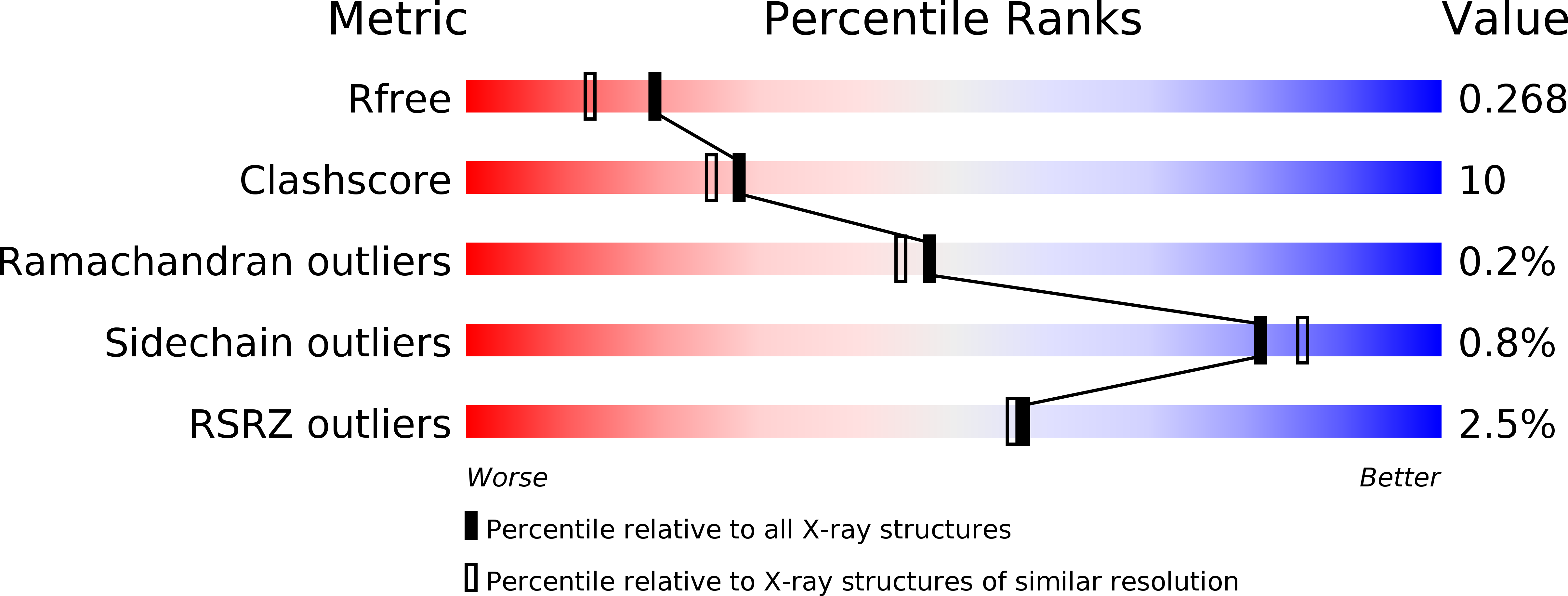
Resolution:
2.00 Å
R-Value Free:
0.26
R-Value Work:
0.21
R-Value Observed:
0.21
Space Group:
P 1 21 1
Macromolecular Entities
Polymer Type:polypeptide(L)
Molecule:Ankyrin-1
Gene (Uniprot):ANK1
Chain IDs:A, B, C, D
Chain Length:161
Number of Molecules:4
Biological Source:Homo sapiens
Ligand Molecules
Primary Citation
Structures of the spectrin-ankyrin interaction binding domains.
Blood 113 5385 5393 (2009)
PMID: 19141864 DOI: 10.1182/blood-2008-10-184358

Abstact

As key components of the erythrocyte membrane skeleton, spectrin and ankyrin specifically interact to tether the spectrin cytoskeleton to the cell membrane. The structure of the spectrin binding domain of ankyrin and the ankyrin binding domain of spectrin have been solved to elucidate the structural basis for ankyrin-spectrin recognition. The structure of repeats 14 and 15 of spectrin shows that these repeats are similar to all other spectrin repeats. One feature that could account for the preference of ankyrin for these repeats is the presence of a conserved, negatively charged patch on one side of repeat 14. The structure of the ankyrin ZU5 domain shows a novel structure containing a beta core. The structure reveals that the canonical ZU5 consensus sequence is likely to be missing an important region that codes for a beta strand that forms part of the core of the domain. In addition, a positively charged region is suggestive of a binding surface for the negatively charged spectrin repeat 14. Previously reported mutants of ankyrin that map to this region lie mostly on the surface of the protein, although at least one is likely to be part of the core.

Legend

Protein

Chemical

Disease

Primary Citation of related structures