
Deposition Date
2008-10-31
Release Date
2009-10-20
Last Version Date
2023-11-01
Entry Detail
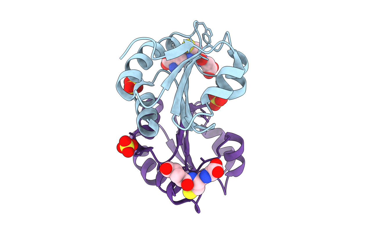
PDB ID:
3F3R
Keywords:
Title:
Crystal structure of yeast Thioredoxin1-glutathione mixed disulfide complex
Biological Source:
Source Organism(s):
Saccharomyces cerevisiae (Taxon ID: 4932)
Expression System(s):
Method Details:
Experimental Method:
Resolution:
1.80 Å
R-Value Free:
0.21
R-Value Work:
0.18
R-Value Observed:
0.18
Space Group:
P 1
