Deposition Date
2008-10-31
Release Date
2009-10-20
Last Version Date
2023-11-01
Entry Detail
PDB ID:
3F3M
Keywords:
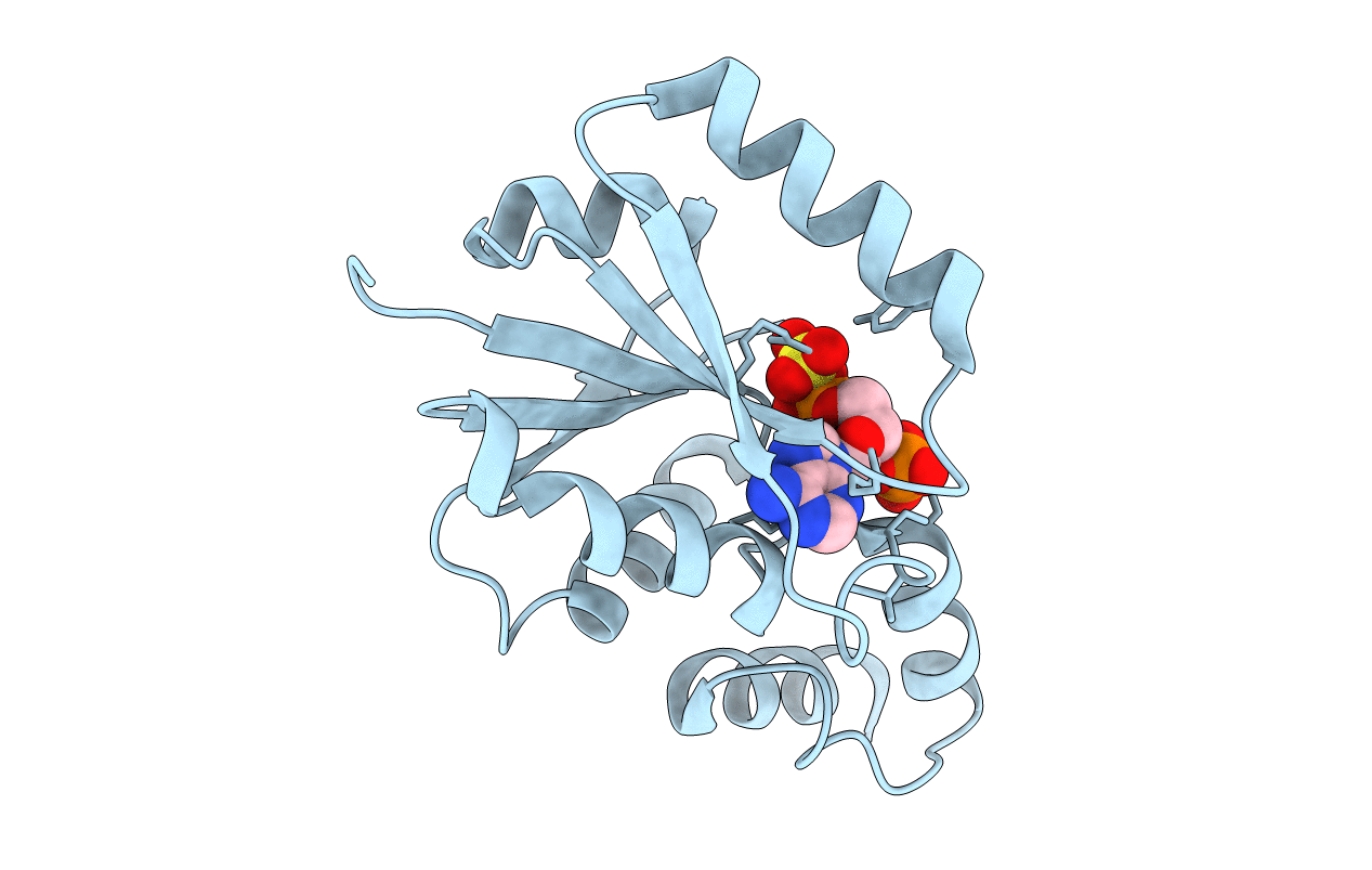
Title:
Six Crystal Structures of Two Phosphopantetheine Adenylyltransferases Reveal an Alternative Ligand Binding Mode and an Associated Structural Change
Biological Source:
Source Organism(s):
Staphylococcus aureus (Taxon ID: 1280)
Expression System(s):
Method Details:
Experimental Method:
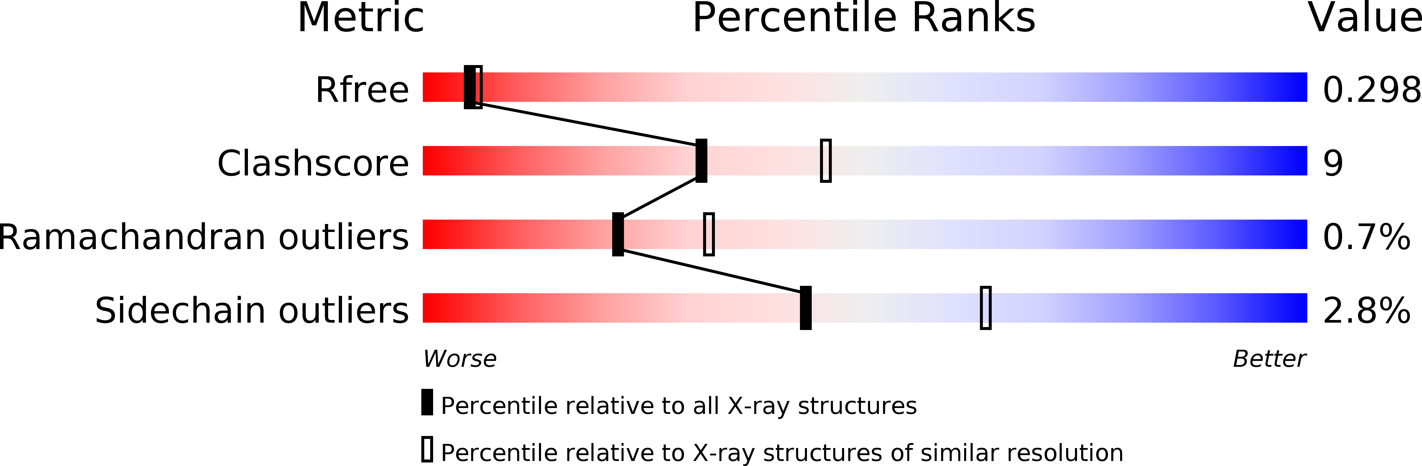
Resolution:
2.40 Å
R-Value Free:
0.25
R-Value Work:
0.21
Space Group:
P 63 2 2