Deposition Date
2008-10-30
Release Date
2009-10-13
Last Version Date
2023-12-27
Entry Detail
PDB ID:
3F31
Keywords:
Title:
Crystal Structure of the N-terminal region of AlphaII-spectrin Tetramerization Domain
Biological Source:
Source Organism(s):
Homo sapiens (Taxon ID: 9606)
Expression System(s):
Method Details:
Experimental Method:
Resolution:
2.30 Å
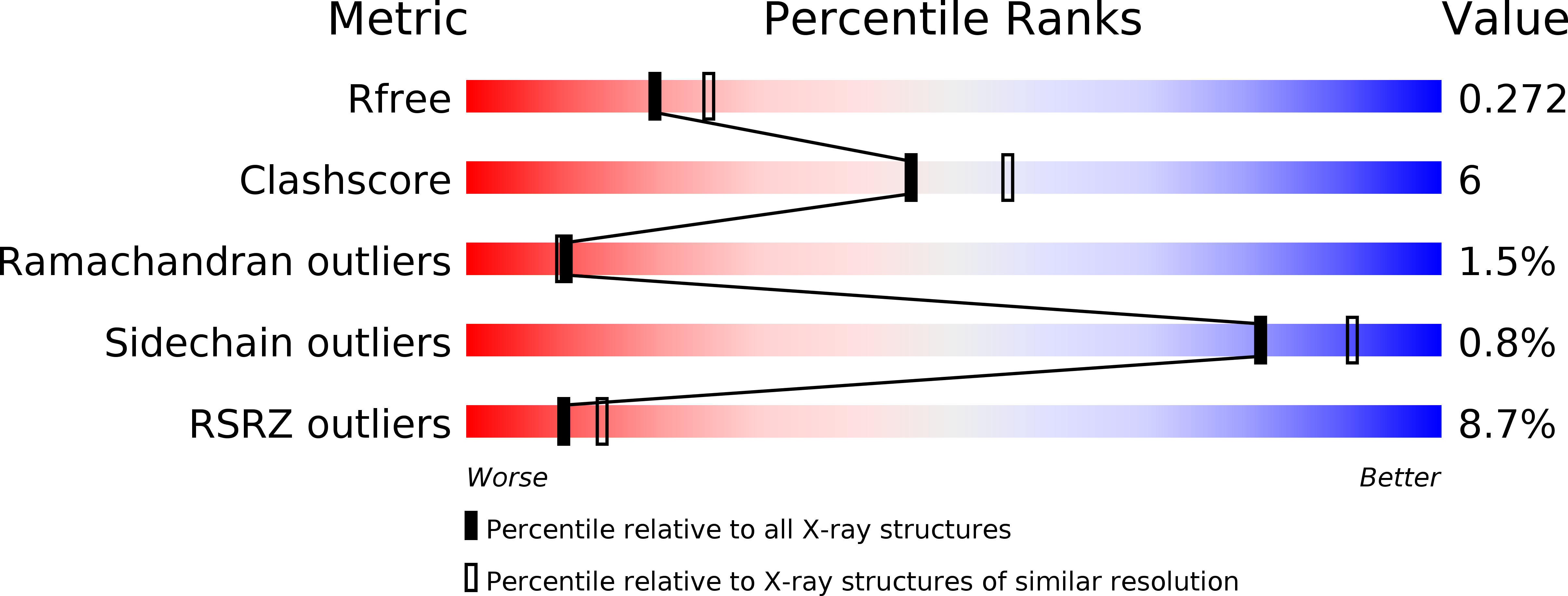
R-Value Free:
0.27
R-Value Work:
0.21
R-Value Observed:
0.22
Space Group:
P 21 21 21