3F2E image
Deposition Date 2008-10-29
Release Date 2009-04-21
Last Version Date 2024-04-03
Entry Detail
PDB ID:
3F2E
Keywords:
Title:
Crystal structure of Yellowstone SIRV coat protein C-terminus
Biological Source:
Host Organism:
Method Details:
Experimental Method:
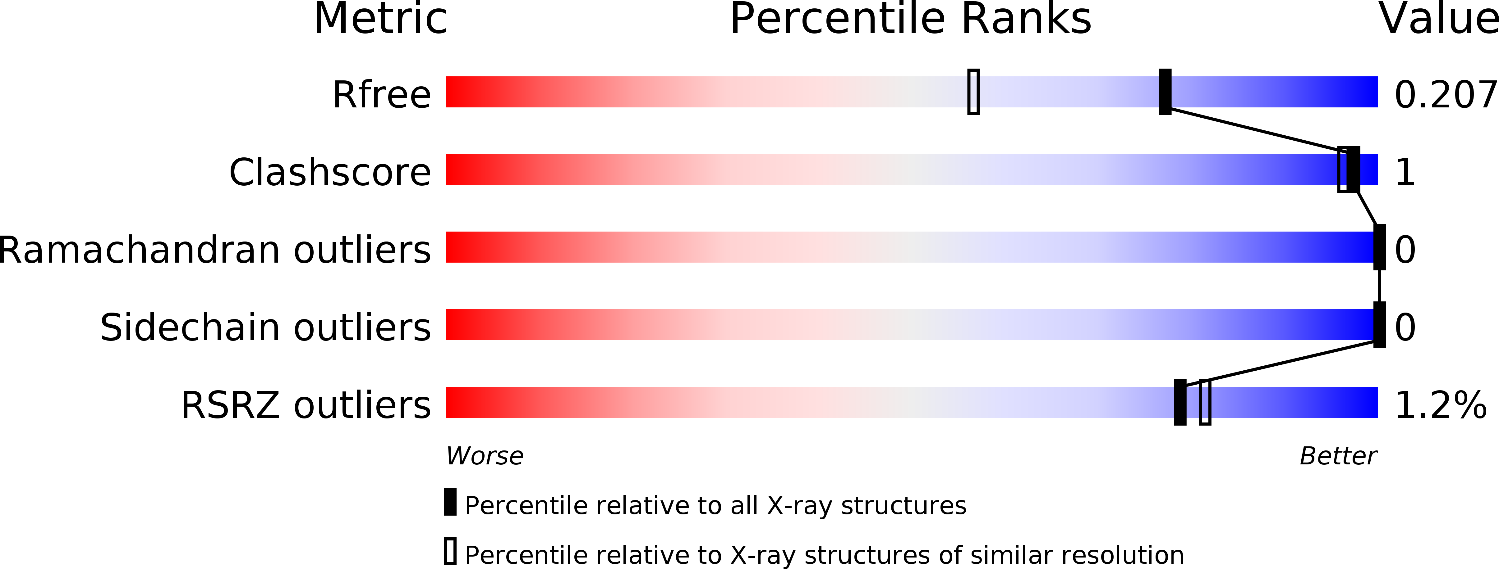
Resolution:
1.67 Å
R-Value Free:
0.20
R-Value Work:
0.18
R-Value Observed:
0.18
Space Group:
P 43 21 2
Macromolecular Entities
Structures with similar UniProt ID
Protein Blast
Polymer Type:polypeptide(L)
Molecule:SIRV coat protein
Chain IDs:A
Chain Length:100
Number of Molecules:1
Biological Source:Sulfolobus islandicus rudivirus 1 variant YNP
Ligand Molecules
Primary Citation
Synergy of NMR, computation, and X-ray crystallography for structural biology.
Structure 17 499 507 (2009)
PMID: 19368883 DOI: 10.1016/j.str.2009.03.001

Abstact

NMR spectroscopy and X-ray crystallography are currently the two most widely applied methods for the determination of macromolecular structures at high resolution. More recently, significant advances have been made in algorithms for the de novo prediction of protein structure, and, in favorable cases, the predicted models agree extremely well with experimentally determined structures. Here, we demonstrate a synergistic combination of NMR spectroscopy, de novo structure prediction, and X-ray crystallography in an effective overall strategy for rapidly determining the structure of the coat protein C-terminal domain from the Sulfolobus islandicus rod-shaped virus (SIRV). This approach takes advantage of the most accessible aspects of each structural technique and may be widely applicable for structure determination.

Legend

Protein

Chemical

Disease

Primary Citation of related structures
Feedback Form
Name
Email
Institute
Feedback