3F1S image
Deposition Date 2008-10-28
Release Date 2009-06-30
Last Version Date 2024-10-30
Entry Detail
PDB ID:
3F1S
Title:
Crystal structure of Protein Z complexed with protein Z-dependent inhibitor
Biological Source:
Source Organism:
Homo sapiens (Taxon ID: 9606)
Host Organism:
Method Details:
Experimental Method:
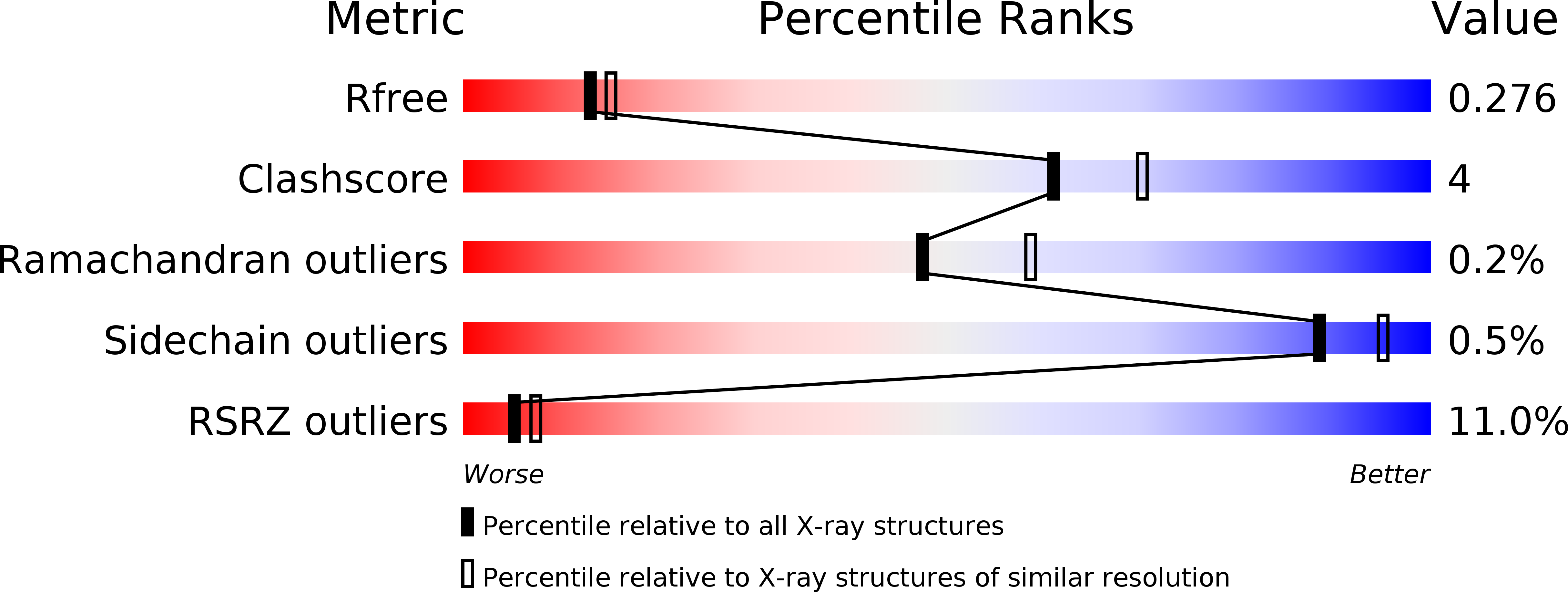
Resolution:
2.30 Å
R-Value Free:
0.27
R-Value Work:
0.22
R-Value Observed:
0.22
Space Group:
P 21 21 21
Macromolecular Entities
Structures with similar UniProt ID
Protein Blast
Polymer Type:polypeptide(L)
Molecule:Protein Z-dependent protease inhibitor
Gene (Uniprot):SERPINA10
Chain IDs:A
Chain Length:385
Number of Molecules:1
Biological Source:Homo sapiens
Structures with similar UniProt ID
Protein Blast
Polymer Type:polypeptide(L)
Molecule:Vitamin K-dependent protein Z
Gene (Uniprot):PROZ
Chain IDs:B
Chain Length:283
Number of Molecules:1
Biological Source:Homo sapiens
Modified Residue
Compound ID Chain ID Parent Comp ID Details 2D Image
ASN B ASN GLYCOSYLATION SITE
Primary Citation
Crystal structure of protein Z-dependent inhibitor complex shows how protein Z functions as a cofactor in the membrane inhibition of factor X.
Blood ? ? ? (2009)
PMID: 19528533 DOI: 10.1182/blood-2009-04-210021

Abstact

Protein Z (PZ) binds to PZ-dependent inhibitor (ZPI) and accelerates the inhibition of the coagulation protease, activated factor X (FXa), in the presence of phospholipids and Ca2+. A 2.3A resolution crystal structure of PZ complexed with ZPI shows that ZPI is a typical serine protease inhibitor and that PZ has a serine protease fold with distorted oxyanion hole and S1 pocket. The 2 molecules bind with fully complementary surfaces spanning over 2400A(2) and involving extensive ionic and hydrophobic interactions. ZPI has an unusual shutter region with a negatively charged residue buried within the hydrophobic core of the molecule. This unique Asp(213) is critical in maintaining the balanced metastability required for optimal protease inhibition, especially when PZ is bound, with its replacement with Asn resulting in increased thermal stability, but decreased efficiency of protease inhibition. The structure of ZPI shows negatively and positively charged surfaces on top of the molecule, in keeping with mutagenesis studies in this work indicating exosite interactions with FXa when it docks on top of ZPI. As modeled in this study, the gamma-carboxy-glutamic acid-containing domains of PZ and FXa enable them to bind to the same phospholipid surfaces on platelet and other membranes, with optimal proximity for the inhibition of FXa by the complexed ZPI.

Legend

Protein

Chemical

Disease

Primary Citation of related structures