3EYX image
Deposition Date 2008-10-22
Release Date 2009-09-15
Last Version Date 2023-11-01
Entry Detail
PDB ID:
3EYX
Keywords:
Title:
Crystal structure of Carbonic Anhydrase Nce103 from Saccharomyces cerevisiae
Biological Source:
Source Organism:
Host Organism:
Method Details:
Experimental Method:
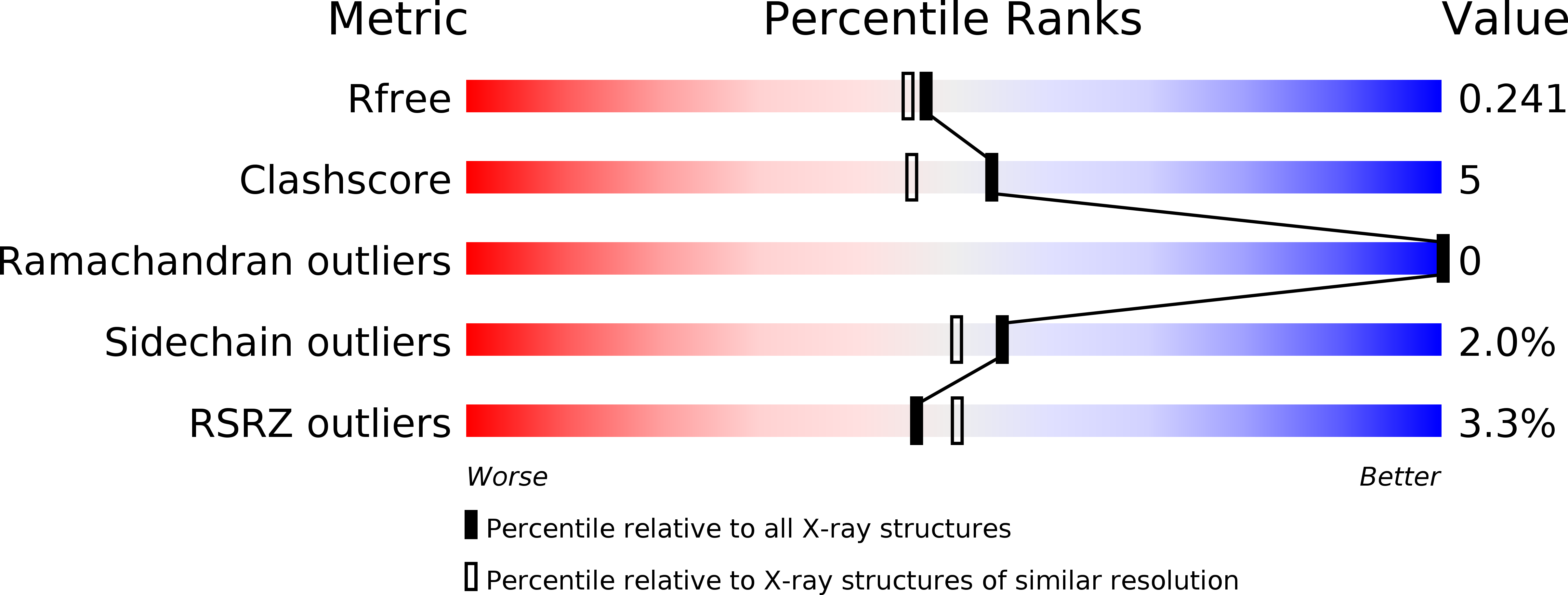
Resolution:
2.04 Å
R-Value Free:
0.24
R-Value Work:
0.19
R-Value Observed:
0.19
Space Group:
C 2 2 21
Macromolecular Entities
Structures with similar UniProt ID
Protein Blast
Polymer Type:polypeptide(L)
Molecule:Carbonic anhydrase
Gene (Uniprot):NCE103
Chain IDs:A, B
Chain Length:216
Number of Molecules:2
Biological Source:Saccharomyces cerevisiae
Primary Citation
Structural insights into the substrate tunnel of Saccharomyces cerevisiae carbonic anhydrase Nce103.
Bmc Struct.Biol. 9 67 67 (2009)
PMID: 19852838 DOI: 10.1186/1472-6807-9-67

Abstact

BACKGROUND The carbonic anhydrases (CAs) are involved in inorganic carbon utilization. They have been classified into six evolutionary and structural families: alpha-, beta-, gamma-, delta-, epsilon-, zeta- CAs, with beta-CAs present in higher plants, algae and prokaryotes. The yeast Saccharomyces cerevisiae encodes a single copy of beta-CA Nce103/YNL036W. RESULTS We determined the crystal structure of Nce103 in complex with a substrate analog at 2.04 A resolution. It assembles as a homodimer, with the active site located at the interface between two monomers. At the bottom of the substrate pocket, a zinc ion is coordinated by the three highly conserved residues Cys57, His112 and Cys115 in addition to a water molecule. Residues Asp59, Arg61, Gly111, Leu102, Val80, Phe75 and Phe97 form a tunnel to the bottom of the active site which is occupied by a molecule of the substrate analog acetate. Activity assays of full length and two truncated versions of Nce103 indicated that the N-terminal arm is indispensable. CONCLUSION The quaternary structure of Nce103 resembles the typical plant type beta-CAs of known structure, with an N-terminal arm indispensable for the enzymatic activity. Comparative structure analysis enables us to draw a possible tunnel for the substrate to access the active site which is located at the bottom of a funnel-shaped substrate pocket.

Legend

Protein

Chemical

Disease

Primary Citation of related structures
Feedback Form
Name
Email
Institute
Feedback