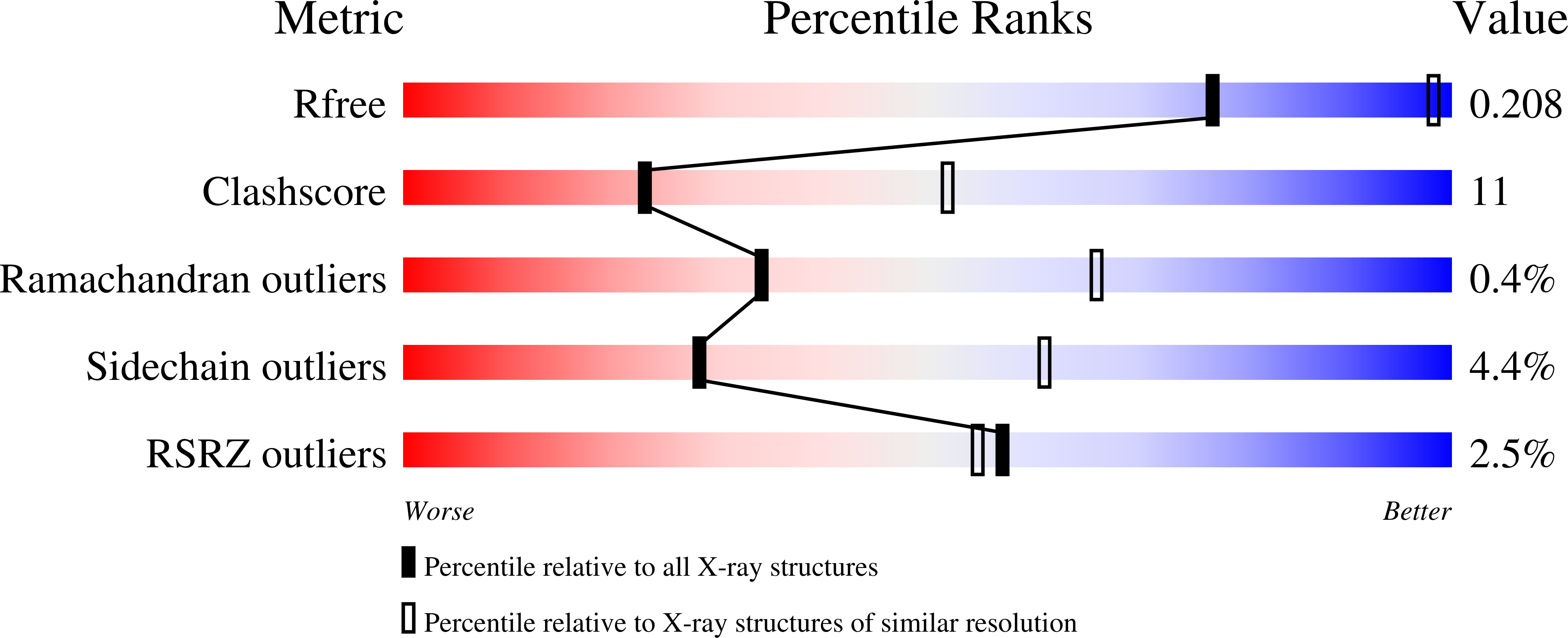
3EY9 image
Deposition Date 2008-10-20
Release Date 2008-11-04
Last Version Date 2023-11-01
Entry Detail
PDB ID:
3EY9
Keywords:
Title:
Structural basis for membrane binding and catalytic activation of the peripheral membrane enzyme pyruvate oxidase from Escherichia coli
Biological Source:
Source Organism:
Escherichia coli (Taxon ID: 562)
Host Organism:
Method Details:
Experimental Method:
Resolution:
2.90 Å
R-Value Free:
0.21
R-Value Work:
0.18
R-Value Observed:
0.18
Space Group:
P 43 21 2
Macromolecular Entities
Structures with similar UniProt ID
Protein Blast
Polymer Type:polypeptide(L)
Molecule:Pyruvate dehydrogenase [cytochrome]
Gene (Uniprot):poxB
Chain IDs:A, B
Chain Length:572
Number of Molecules:2
Biological Source:Escherichia coli
Primary Citation
Structural basis for membrane binding and catalytic activation of the peripheral membrane enzyme pyruvate oxidase from Escherichia coli.
Proc.Natl.Acad.Sci.USA 105 17390 17395 (2008)
PMID: 18988747 DOI: 10.1073/pnas.0805027105

Abstact

The thiamin- and flavin-dependent peripheral membrane enzyme pyruvate oxidase from E. coli catalyzes the oxidative decarboxylation of the central metabolite pyruvate to CO(2) and acetate. Concomitant reduction of the enzyme-bound flavin triggers membrane binding of the C terminus and shuttling of 2 electrons to ubiquinone 8, a membrane-bound mobile carrier of the electron transport chain. Binding to the membrane in vivo or limited proteolysis in vitro stimulate the catalytic proficiency by 2 orders of magnitude. The molecular mechanisms by which membrane binding and activation are governed have remained enigmatic. Here, we present the X-ray crystal structures of the full-length enzyme and a proteolytically activated truncation variant lacking the last 23 C-terminal residues inferred as important in membrane binding. In conjunction with spectroscopic results, the structural data pinpoint a conformational rearrangement upon activation that exposes the autoinhibitory C terminus, thereby freeing the active site. In the activated enzyme, Phe-465 swings into the active site and wires both cofactors for efficient electron transfer. The isolated C terminus, which has no intrinsic helix propensity, folds into a helical structure in the presence of micelles.

Legend

Protein

Chemical

Disease

Primary Citation of related structures