3EXS image
Deposition Date 2008-10-17
Release Date 2009-08-25
Last Version Date 2023-11-01
Entry Detail
PDB ID:
3EXS
Keywords:
Title:
Crystal structure of KGPDC from Streptococcus mutans in complex with D-R5P
Biological Source:
Source Organism:
Host Organism:
Method Details:
Experimental Method:
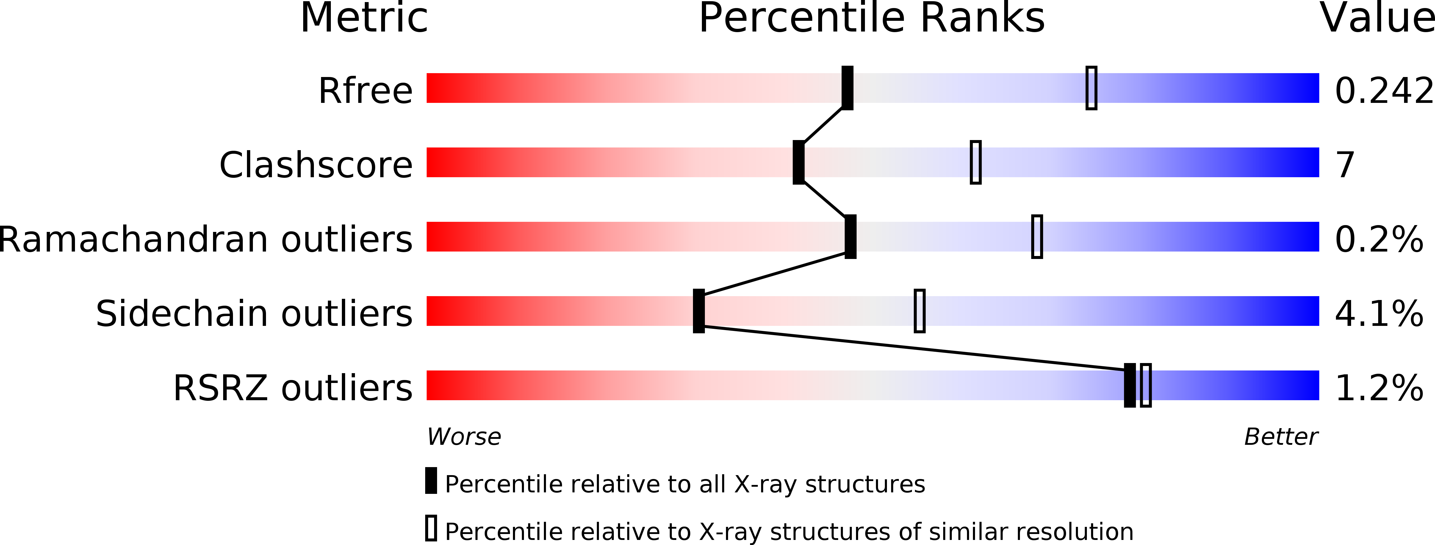
Resolution:
2.50 Å
R-Value Free:
0.24
R-Value Work:
0.18
R-Value Observed:
0.18
Space Group:
P 21 21 21
Macromolecular Entities
Polymer Type:polypeptide(L)
Molecule:RmpD (Hexulose-6-phosphate synthase)
Gene (Uniprot):rmpD
Chain IDs:A, B, C, D
Chain Length:221
Number of Molecules:4
Biological Source:Streptococcus mutans
Ligand Molecules
Primary Citation
Open-closed conformational change revealed by the crystal structures of 3-keto-L-gulonate 6-phosphate decarboxylase from Streptococcus mutans
Biochem.Biophys.Res.Commun. 381 429 433 (2009)
PMID: 19222992 DOI: 10.1016/j.bbrc.2009.02.049

Abstact

The 3-keto-L-gulonate 6-phosphate decarboxylase (KGPDC) catalyses the decarboxylation of 3-keto-L-gulonate 6-phosphate to L-xylulose in the presence of magnesium ions. The enzyme is involved in L-ascorbate metabolism and plays an essential role in the pathway of glucuronate interconversion. Crystal structures of Streptococcus mutans KGPDC were determined in the absence and presence of the product analog D-ribulose 5-phosphate. We have observed an 8 A alphaB-helix movement and other structural rearrangements around the active site between the apo-structures and product analog bound structure. These drastic conformational changes upon ligand binding are the first observation of this kind for the KGPDC family. The flexibilities of both the alpha-helix lid and the side chains of Arg144 and Arg197 are associated with substrate binding and product releasing. The open-closed conformational changes of the active site, through the movements of the alpha-helix lid and the arginine residues are important for substrate binding and catalysis.

Legend

Protein

Chemical

Disease

Primary Citation of related structures