3EXJ image
Deposition Date 2008-10-16
Release Date 2008-12-16
Last Version Date 2023-12-27
Entry Detail
PDB ID:
3EXJ
Title:
Crystal Structure of a p53 Core Tetramer Bound to DNA
Biological Source:
Source Organism:
Mus musculus (Taxon ID: 10090)
Host Organism:
Method Details:
Experimental Method:
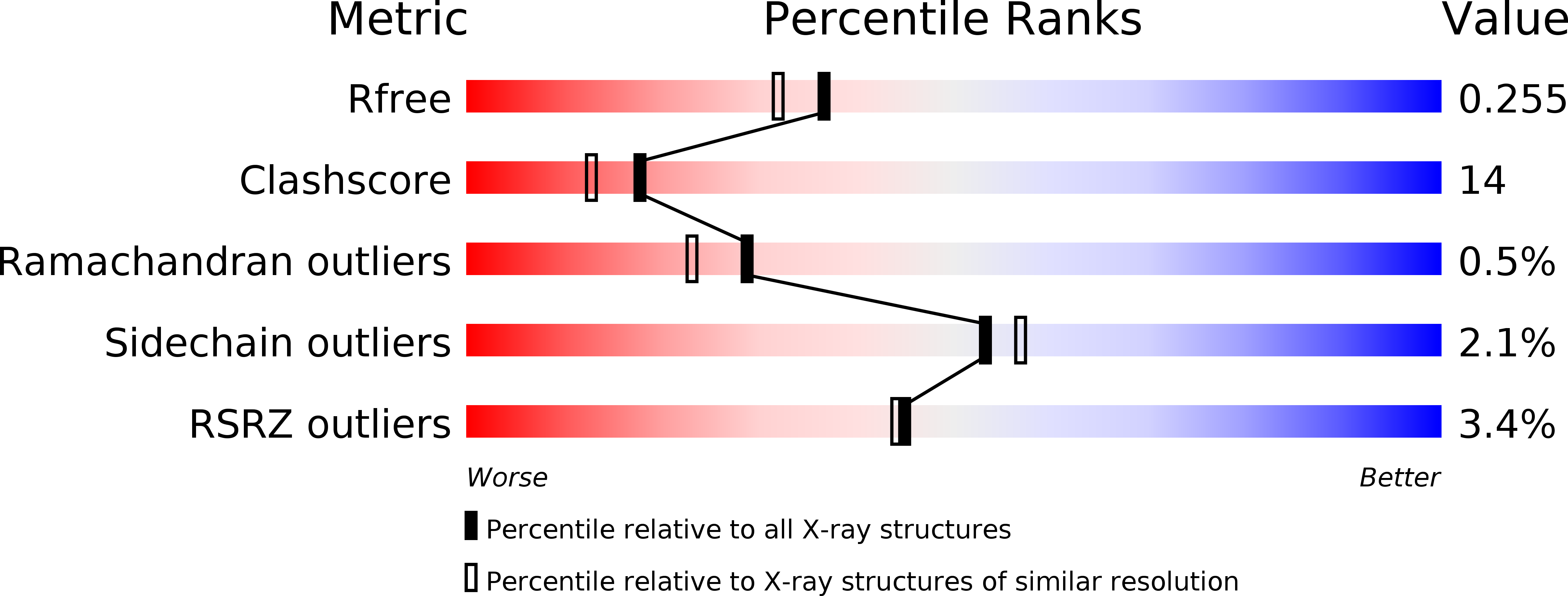
Resolution:
2.00 Å
R-Value Free:
0.26
R-Value Work:
0.22
Space Group:
C 1 2 1
Macromolecular Entities
Polymer Type:polypeptide(L)
Molecule:mouse p53 core domain
Gene (Uniprot):Tp53
Chain IDs:A, B
Chain Length:197
Number of Molecules:2
Biological Source:Mus musculus
Primary Citation
Crystal structure of a p53 core tetramer bound to DNA.
Oncogene 28 325 333 (2009)
PMID: 18978813 DOI: 10.1038/onc.2008.400

Abstact

The tumor suppressor p53 regulates downstream genes in response to many cellular stresses and is frequently mutated in human cancers. Here, we report the use of a crosslinking strategy to trap a tetrameric p53 DNA-binding domain (p53DBD) bound to DNA and the X-ray crystal structure of the protein/DNA complex. The structure reveals that two p53DBD dimers bind to B form DNA with no relative twist and that a p53 tetramer can bind to DNA without introducing significant DNA bending. The numerous dimer-dimer interactions involve several strictly conserved residues, thus suggesting a molecular basis for p53DBD-DNA binding cooperativity. Surface residue conservation of the p53DBD tetramer bound to DNA highlights possible regions of other p53 domain or p53 cofactor interactions.

Legend

Protein

Chemical

Disease

Primary Citation of related structures