Deposition Date
2008-10-15
Release Date
2009-03-24
Last Version Date
2023-12-27
Entry Detail
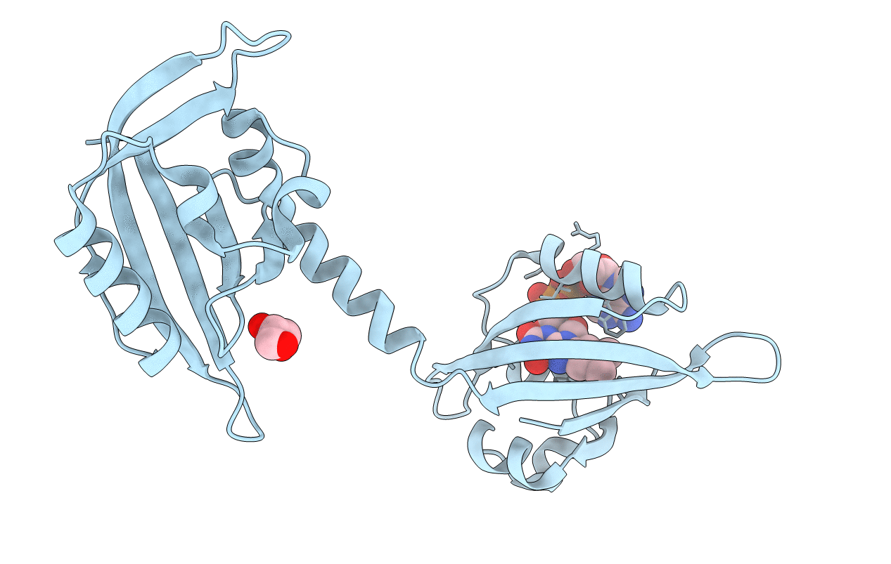
PDB ID:
3EWK
Keywords:
Title:
Structure of the redox sensor domain of Methylococcus capsulatus (Bath) MmoS
Biological Source:
Source Organism(s):
Methylococcus capsulatus (Taxon ID: 414)
Expression System(s):
Method Details:
Experimental Method:
Resolution:
2.34 Å
R-Value Free:
0.28
R-Value Work:
0.23
R-Value Observed:
0.24
Space Group:
P 4 21 2