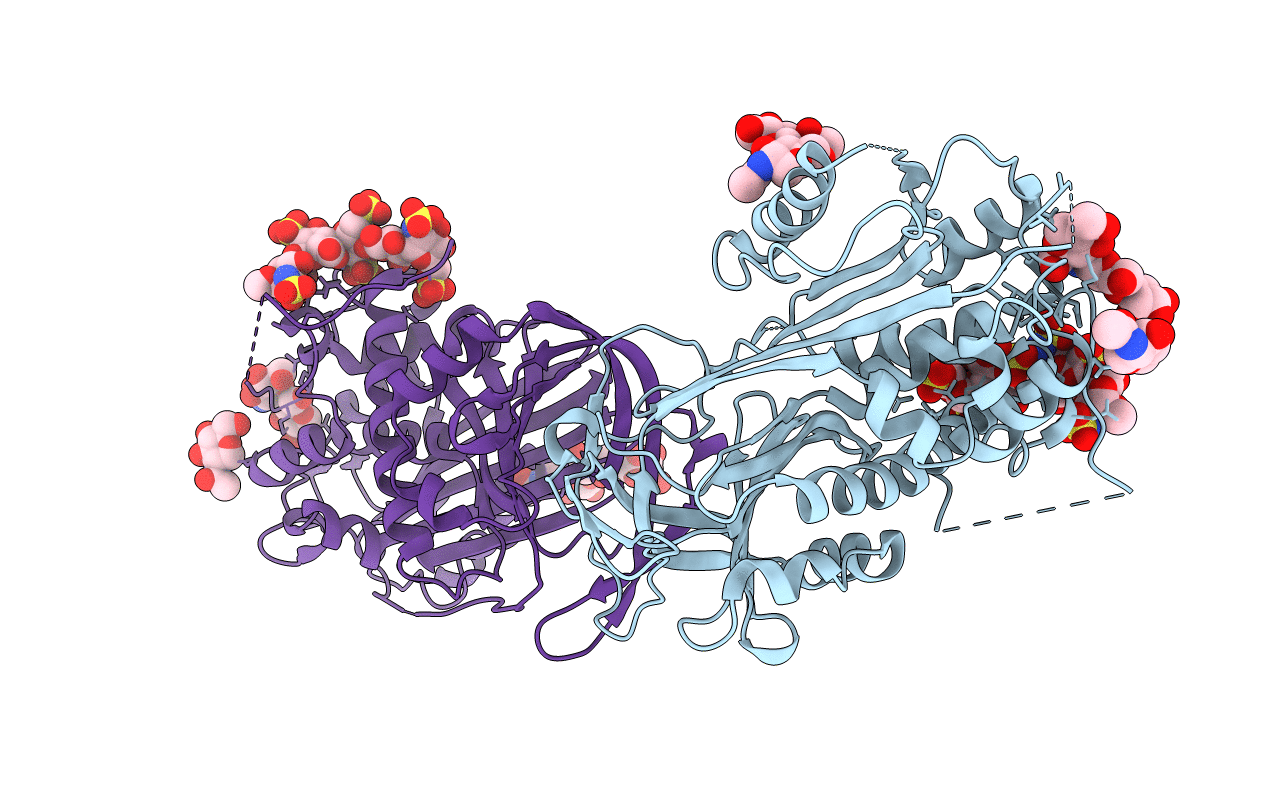
3EVJ image
Deposition Date 2008-10-13
Release Date 2008-10-21
Last Version Date 2024-11-20
Entry Detail
PDB ID:
3EVJ
Title:
Intermediate structure of antithrombin bound to the natural pentasaccharide
Biological Source:
Source Organism:
Homo sapiens (Taxon ID: 9606)
Method Details:
Experimental Method:
Resolution:
3.00 Å
R-Value Free:
0.28
R-Value Work:
0.23
R-Value Observed:
0.23
Space Group:
P 1 21 1
Macromolecular Entities
Polymer Type:polypeptide(L)
Molecule:Antithrombin-III
Gene (Uniprot):SERPINC1
Chain IDs:A (auth: I), B (auth: L)
Chain Length:432
Number of Molecules:2
Biological Source:Homo sapiens
Modified Residue
Compound ID Chain ID Parent Comp ID Details 2D Image
ASN A ASN GLYCOSYLATION SITE
Ligand Molecules
Peptide-like Molecules
PRD_900028
Primary Citation
The critical role of hinge-region expulsion in the induced-fit heparin binding mechanism of antithrombin.
J. Mol. Biol. 386 1278 1289 (2009)
PMID: 19452598

Abstact

Antithrombin (AT) is the most important inhibitor of coagulation proteases. Its activity is stimulated by glycosaminoglycans, such as heparin, through allosteric and template mechanisms. AT utilises an induced-fit mechanism to bind with high affinity to a pentasaccharide sequence found in about one-third of heparin chains. The conformational changes behind this mechanism have been characterised by several crystal structures of AT in the absence and in the presence of pentasaccharide. Pentasaccharide binding ultimately results in a conformational change that improves affinity by about 1000-fold. Crystal structures show several differences, including the expulsion of the hinge region of the reactive centre loop from beta-sheet A, which is known to be critical for the allosteric activation of AT. Here, we present data that reveal an energetically distinct intermediate on the path to full activation where the majority of conformational changes have already occurred. A crystal structure of this intermediate shows that the hinge region is in a native-like state in spite of having the pentasaccharide bound in the normal fashion. We engineered a disulfide bond to lock the hinge in its native position to determine the energetic contributions of the initial and final conformational events. Approximately 60% of the free-energy contribution of conformational change is provided by the final step of hinge-region expulsion and subsequent closure of the main beta-sheet A. A new analysis of the individual structural changes provides a plausible mechanism for propagation of conformational change from the heparin binding site to the remote hinge region in beta-sheet A.

Legend

Protein

Chemical

Disease

Primary Citation of related structures