3ETC image
Deposition Date 2008-10-07
Release Date 2009-07-07
Last Version Date 2023-12-27
Entry Detail
PDB ID:
3ETC
Keywords:
Title:
2.1 A structure of acyl-adenylate synthetase from Methanosarcina acetivorans containing a link between Lys256 and Cys298
Biological Source:
Source Organism(s):
Expression System(s):
Method Details:
Experimental Method:
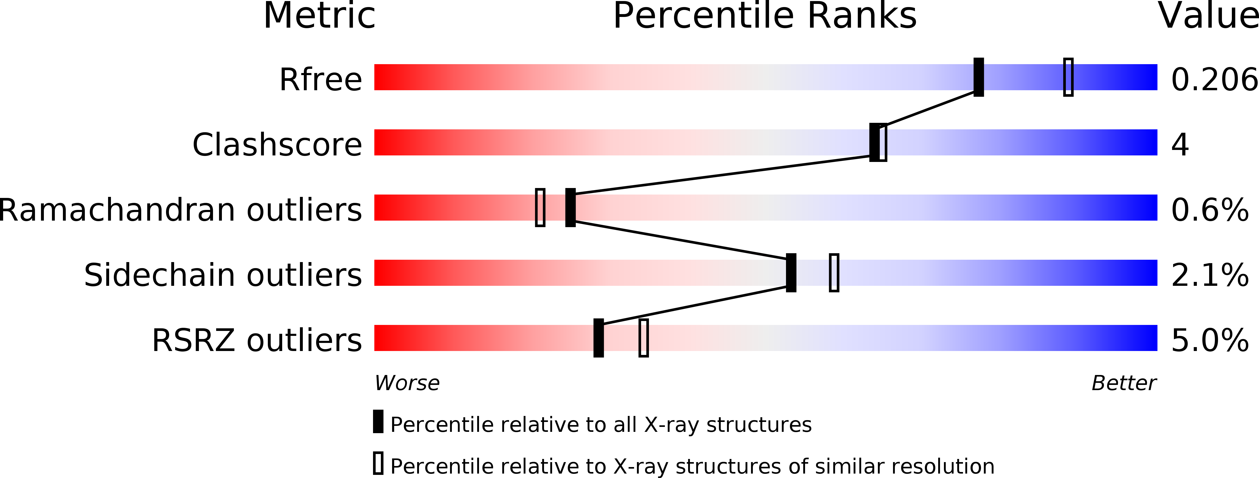
Resolution:
2.10 Å
R-Value Free:
0.20
R-Value Work:
0.17
R-Value Observed:
0.17
Space Group:
P 21 21 21
Macromolecular Entities
Structures with similar UniProt ID
Protein Blast
Polymer Type:polypeptide(L)
Molecule:AMP-binding protein
Gene (Uniprot):MA_2912
Chain IDs:A, B
Chain Length:580
Number of Molecules:2
Biological Source:Methanosarcina acetivorans
Primary Citation
The 2.1 A crystal structure of an acyl-CoA synthetase from Methanosarcina acetivorans reveals an alternate acyl-binding pocket for small branched acyl substrates.
Proteins 77 685 698 (2009)
PMID: 19544569 DOI: 10.1002/prot.22482

Abstact

The acyl-AMP forming family of adenylating enzymes catalyze two-step reactions to activate a carboxylate with the chemical energy derived from ATP hydrolysis. X-ray crystal structures have been determined for multiple members of this family and, together with biochemical studies, provide insights into the active site and catalytic mechanisms used by these enzymes. These studies have shown that the enzymes use a domain rotation of 140 degrees to reconfigure a single active site to catalyze the two partial reactions. We present here the crystal structure of a new medium chain acyl-CoA synthetase from Methanosarcina acetivorans. The binding pocket for the three substrates is analyzed, with many conserved residues present in the AMP binding pocket. The CoA binding pocket is compared to the pockets of both acetyl-CoA synthetase and 4-chlorobenzoate:CoA ligase. Most interestingly, the acyl-binding pocket of the new structure is compared with other acyl- and aryl-CoA synthetases. A comparison of the acyl-binding pocket of the acyl-CoA synthetase from M. acetivorans with other structures identifies a shallow pocket that is used to bind the medium chain carboxylates. These insights emphasize the high sequence and structural diversity among this family in the area of the acyl-binding pocket.

Legend

Protein

Chemical

Disease

Primary Citation of related structures
Feedback Form
Name
Email
Institute
Feedback