3ET6 image
Deposition Date 2008-10-07
Release Date 2008-10-14
Last Version Date 2024-11-20
Entry Detail
PDB ID:
3ET6
Keywords:
Title:
The crystal structure of the catalytic domain of a eukaryotic guanylate cyclase
Biological Source:
Source Organism:
Host Organism:
Method Details:
Experimental Method:
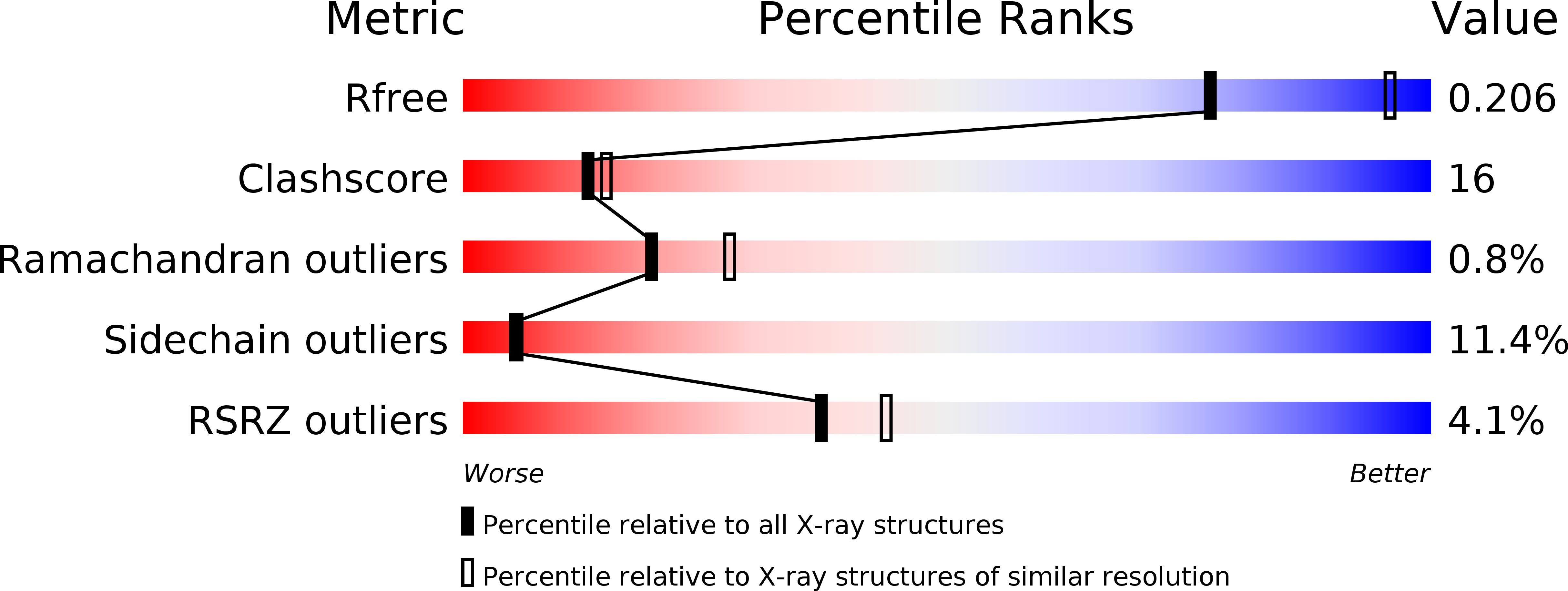
Resolution:
2.55 Å
R-Value Free:
0.21
R-Value Work:
0.17
R-Value Observed:
0.17
Space Group:
P 32 2 1
Macromolecular Entities
Structures with similar UniProt ID
Protein Blast
Polymer Type:polypeptide(L)
Molecule:Soluble guanylyl cyclase beta
Gene (Uniprot):CHLRE_07g320750v5
Chain IDs:A
Chain Length:190
Number of Molecules:1
Biological Source:Chlamydomonas reinhardtii
Structures with similar UniProt ID
Protein Blast
Polymer Type:polypeptide(L)
Molecule:Soluble guanylyl cyclase beta
Gene (Uniprot):CHLRE_07g320750v5
Chain IDs:B
Chain Length:190
Number of Molecules:1
Biological Source:Chlamydomonas reinhardtii
Modified Residue
Compound ID Chain ID Parent Comp ID Details 2D Image
CAS A CYS S-(DIMETHYLARSENIC)CYSTEINE
Ligand Molecules
Primary Citation
The crystal structure of the catalytic domain of a eukaryotic guanylate cyclase.
Bmc Struct.Biol. 8 42 42 (2008)
PMID: 18842118 DOI: 10.1186/1472-6807-8-42

Abstact

BACKGROUND Soluble guanylate cyclases generate cyclic GMP when bound to nitric oxide, thereby linking nitric oxide levels to the control of processes such as vascular homeostasis and neurotransmission. The guanylate cyclase catalytic module, for which no structure has been determined at present, is a class III nucleotide cyclase domain that is also found in mammalian membrane-bound guanylate and adenylate cyclases. RESULTS We have determined the crystal structure of the catalytic domain of a soluble guanylate cyclase from the green algae Chlamydomonas reinhardtii at 2.55 A resolution, and show that it is a dimeric molecule. CONCLUSION Comparison of the structure of the guanylate cyclase domain with the known structures of adenylate cyclases confirms the close similarity in architecture between these two enzymes, as expected from their sequence similarity. The comparison also suggests that the crystallized guanylate cyclase is in an inactive conformation, and the structure provides indications as to how activation might occur. We demonstrate that the two active sites in the dimer exhibit positive cooperativity, with a Hill coefficient of approximately 1.5. Positive cooperativity has also been observed in the homodimeric mammalian membrane-bound guanylate cyclases. The structure described here provides a reliable model for functional analysis of mammalian guanylate cyclases, which are closely related in sequence.

Legend

Protein

Chemical

Disease

Primary Citation of related structures