Deposition Date
2008-10-06
Release Date
2008-10-14
Last Version Date
2023-12-27
Entry Detail
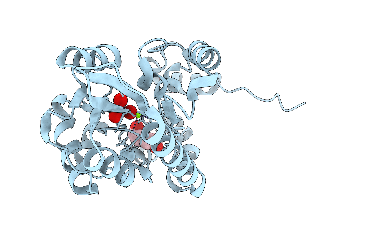
PDB ID:
3ET5
Keywords:
Title:
Structure of Recombinant Haemophilus Influenzae E(P4) Acid Phosphatase Complexed with tungstate
Biological Source:
Source Organism(s):
Haemophilus influenzae (Taxon ID: 281310)
Expression System(s):
Method Details:
Experimental Method:
Resolution:
2.00 Å
R-Value Free:
0.22
R-Value Work:
0.18
R-Value Observed:
0.18
Space Group:
P 42 21 2