Deposition Date
2008-10-06
Release Date
2009-02-17
Last Version Date
2023-12-27
Entry Detail
PDB ID:
3ESL
Keywords:
Title:
Crystal structure of the conserved N-terminal domain of the mitotic checkpoint component BUB1
Biological Source:
Source Organism:
Saccharomyces cerevisiae (Taxon ID: 4932)
Host Organism:
Method Details:
Experimental Method:
Resolution:
1.74 Å
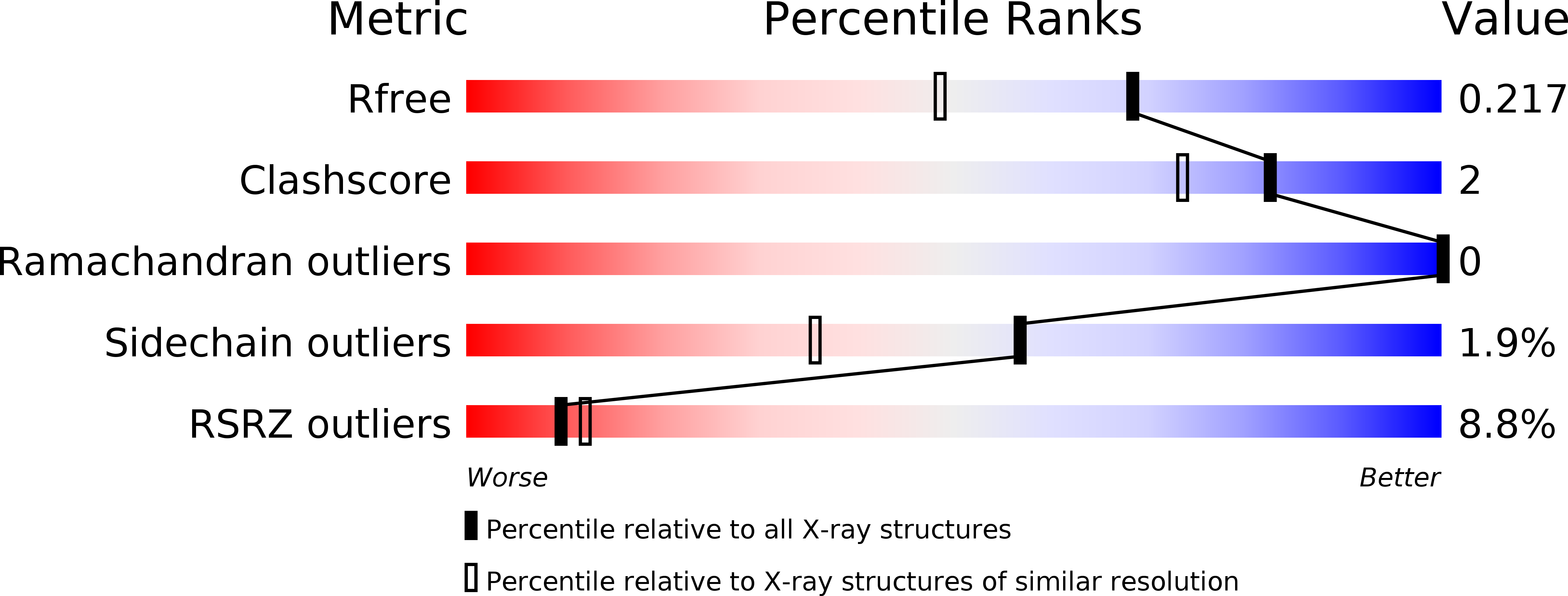
R-Value Free:
0.21
R-Value Work:
0.19
R-Value Observed:
0.19
Space Group:
C 1 2 1