3ESK image
Deposition Date 2008-10-06
Release Date 2009-07-07
Last Version Date 2023-09-06
Entry Detail
PDB ID:
3ESK
Keywords:
Title:
Structure of HOP TPR2A domain in complex with the non-cognate Hsc70 peptide ligand
Biological Source:
Source Organism:
Homo sapiens (Taxon ID: 9606)
Host Organism:
Method Details:
Experimental Method:
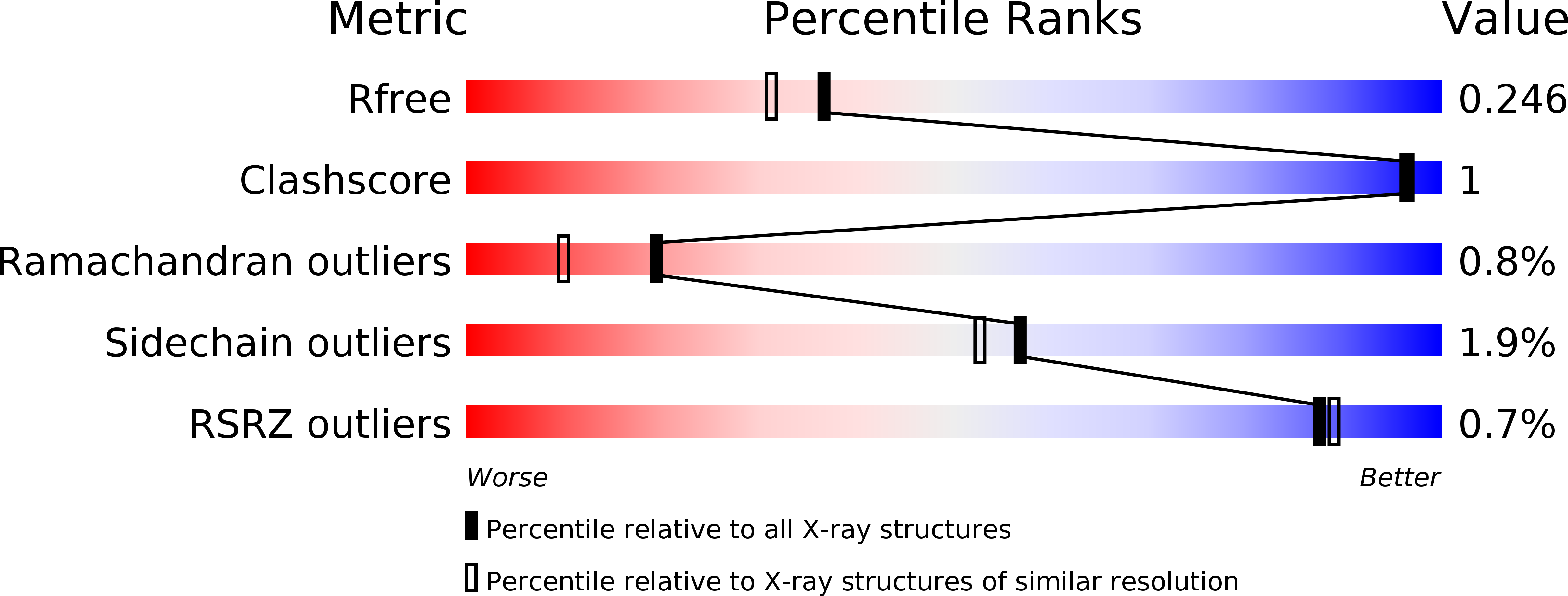
Resolution:
2.05 Å
R-Value Free:
0.24
R-Value Work:
0.17
R-Value Observed:
0.18
Space Group:
C 1 2 1
Macromolecular Entities
Polymer Type:polypeptide(L)
Molecule:Stress-induced-phosphoprotein 1
Gene (Uniprot):STIP1
Chain IDs:A
Chain Length:129
Number of Molecules:1
Biological Source:Homo sapiens
Polymer Type:polypeptide(L)
Molecule:Heat shock cognate 71 kDa protein
Gene (Uniprot):HSPA8
Chain IDs:B
Chain Length:12
Number of Molecules:1
Biological Source:Homo sapiens
Ligand Molecules
Primary Citation
Electrostatic interactions of Hsp-organizing protein tetratricopeptide domains with Hsp70 and Hsp90: computational analysis and protein engineering.
J.Biol.Chem. 284 25364 25374 (2009)
PMID: 19586912 DOI: 10.1074/jbc.M109.033894

Abstact

The Hsp-organizing protein (HOP) binds to the C termini of the chaperones Hsp70 and Hsp90, thus bringing them together so that substrate proteins can be passed from Hsp70 to Hsp90. Because Hsp90 is essential for the correct folding and maturation of many oncogenic proteins, it has become a significant target for anti-cancer drug design. HOP binds to Hsp70 and Hsp90 via two independent tetratricopeptide (TPR) domains, TPR1 and TPR2A, respectively. We have analyzed ligand binding using Poisson-Boltzmann continuum electrostatic calculations, free energy perturbation, molecular dynamics simulations, and site-directed mutagenesis to delineate the contribution of different interactions to the affinity and specificity of the TPR-peptide interactions. We found that continuum electrostatic calculations could be used to guide protein design by removing unfavorable interactions to increase binding affinity, with an 80-fold increase in affinity for TPR2A. Contributions at buried charged residues, however, were better predicted by free energy perturbation calculations. We suggest using a combination of the two approaches for increasing the accuracy of results, with free energy perturbation calculations used only at selected buried residues of the ligand binding pocket. Finally we present the crystal structure of TPR2A in complex with its non-cognate Hsp70 ligand, which provides insight on the origins of specificity in TPR domain-peptide recognition.

Legend

Protein

Chemical

Disease

Primary Citation of related structures