Deposition Date
2008-10-01
Release Date
2009-03-10
Last Version Date
2024-10-30
Entry Detail
PDB ID:
3ERB
Keywords:
Title:
The Crystal Structure of C2b, a Fragment of Complement Component C2 produced during C3-convertase Formation
Biological Source:
Source Organism(s):
Homo sapiens (Taxon ID: 9606)
Expression System(s):
Method Details:
Experimental Method:
Resolution:
1.80 Å
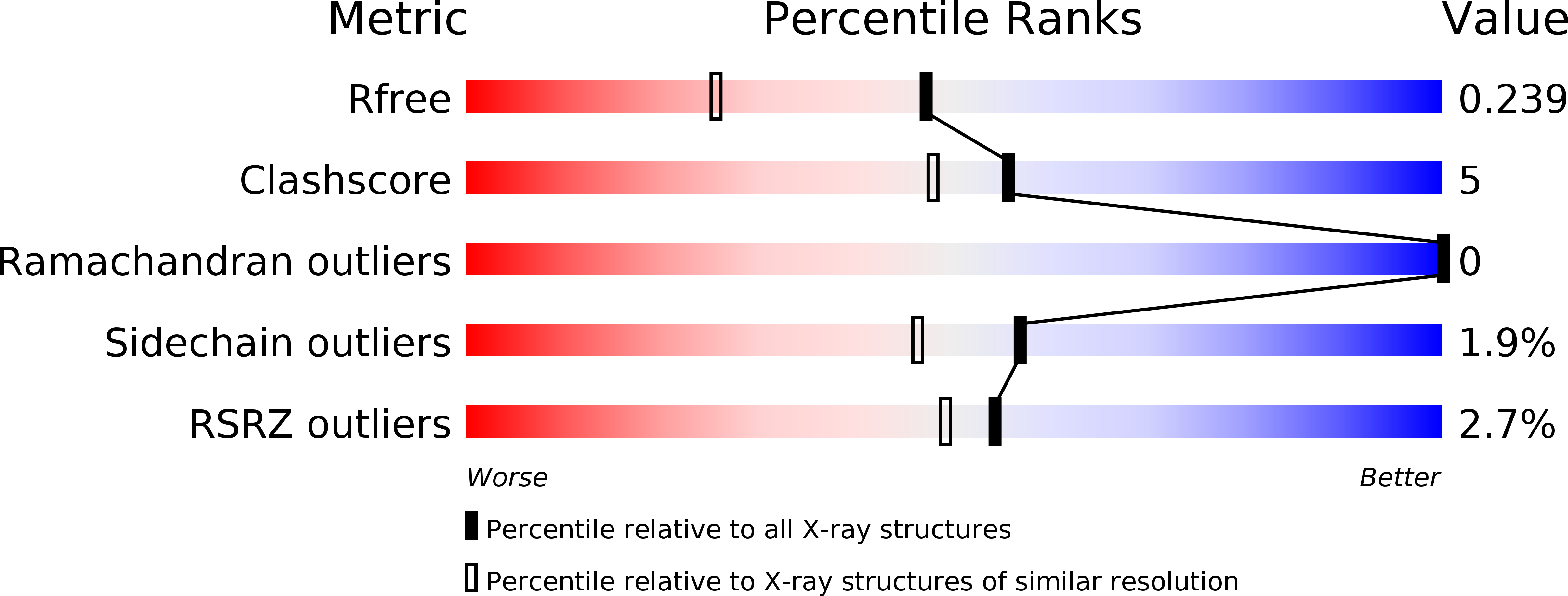
R-Value Free:
0.23
R-Value Work:
0.21
R-Value Observed:
0.21
Space Group:
P 31