3EQV image
Deposition Date 2008-10-01
Release Date 2008-10-21
Last Version Date 2024-10-16
Entry Detail
PDB ID:
3EQV
Title:
Crystal structure of penicillin-binding protein 2 from Neisseria gonorrhoeae containing four mutations associated with penicillin resistance
Biological Source:
Source Organism:
Host Organism:
Method Details:
Experimental Method:
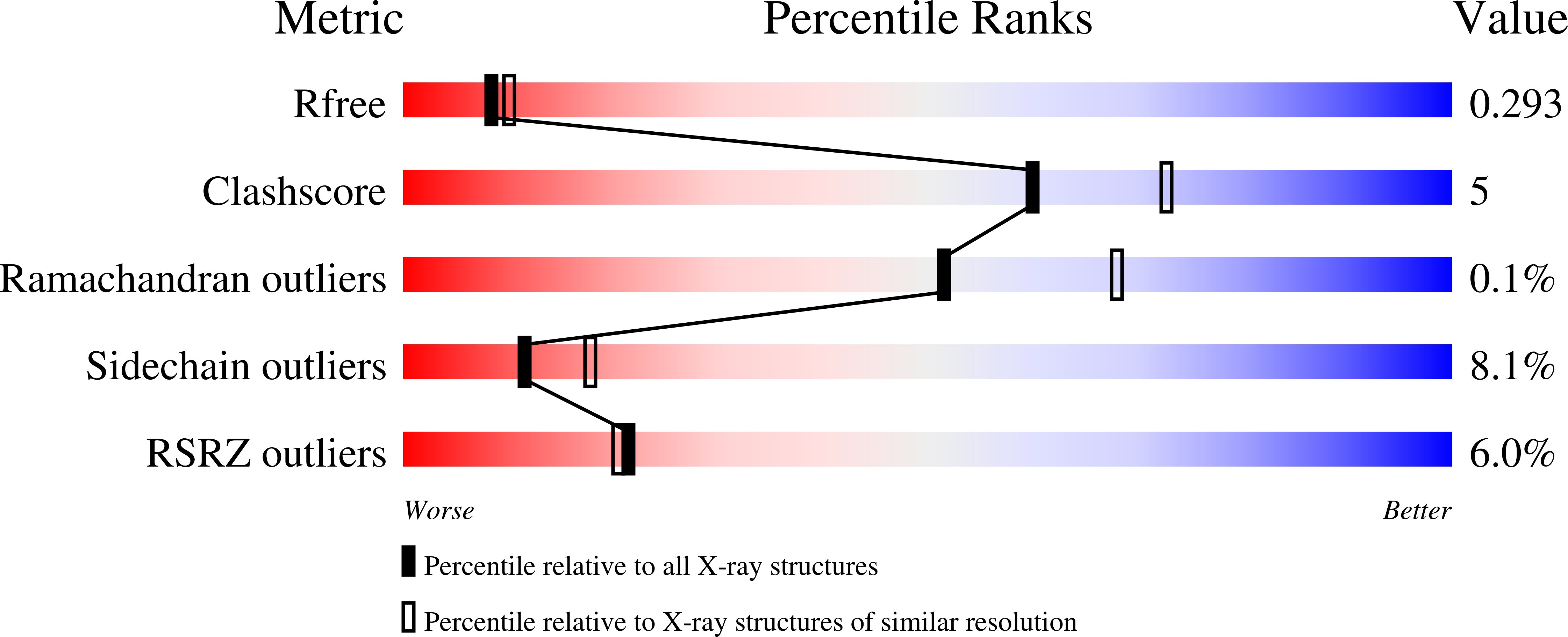
Resolution:
2.40 Å
R-Value Free:
0.25
R-Value Work:
0.21
R-Value Observed:
0.21
Space Group:
P 21 21 21
Macromolecular Entities
Structures with similar UniProt ID
Protein Blast
Polymer Type:polypeptide(L)
Molecule:Penicillin-binding protein 2
Gene (Uniprot):penA
Mutagens:F504L, A510V, A516G, P551S
Chain IDs:A, B
Chain Length:542
Number of Molecules:2
Biological Source:Neisseria gonorrhoeae
Modified Residue
Compound ID Chain ID Parent Comp ID Details 2D Image
SEP A SER PHOSPHOSERINE
Primary Citation
Crystal Structures of Penicillin-binding Protein 2 from Penicillin-susceptible and -resistant Strains of Neisseria gonorrhoeae Reveal an Unexpectedly Subtle Mechanism for Antibiotic Resistance.
J.Biol.Chem. 284 1202 1212 (2009)
PMID: 18986991 DOI: 10.1074/jbc.M805761200

Abstact

Penicillin-binding protein 2 (PBP2) from N. gonorrhoeae is the major molecular target for beta-lactam antibiotics used to treat gonococcal infections. PBP2 from penicillin-resistant strains of N. gonorrhoeae harbors an aspartate insertion after position 345 (Asp-345a) and 4-8 additional mutations, but how these alter the architecture of the protein is unknown. We have determined the crystal structure of PBP2 derived from the penicillin-susceptible strain FA19, which shows that the likely effect of Asp-345a is to alter a hydrogen-bonding network involving Asp-346 and the SXN triad at the active site. We have also solved the crystal structure of PBP2 derived from the penicillin-resistant strain FA6140 that contains four mutations near the C terminus of the protein. Although these mutations lower the second order rate of acylation for penicillin by 5-fold relative to wild type, comparison of the two structures shows only minor structural differences, with the positions of the conserved residues in the active site essentially the same in both. Kinetic analyses indicate that two mutations, P551S and F504L, are mainly responsible for the decrease in acylation rate. Melting curves show that the four mutations lower the thermal stability of the enzyme. Overall, these data suggest that the molecular mechanism underlying antibiotic resistance contributed by the four mutations is subtle and involves a small but measurable disordering of residues in the active site region that either restricts the binding of antibiotic or impedes conformational changes that are required for acylation by beta-lactam antibiotics.

Legend

Protein

Chemical

Disease

Primary Citation of related structures