3EQO image
Deposition Date 2008-10-01
Release Date 2009-02-03
Last Version Date 2024-10-16
Entry Detail
PDB ID:
3EQO
Keywords:
Title:
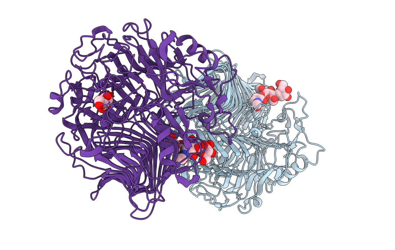
Crystal structure of beta-1,3-glucanase from Phanerochaete chrysosporium (Lam55A) gluconolactone complex
Biological Source:
Source Organism:
Host Organism:
Method Details:
Experimental Method:
Resolution:
2.25 Å
R-Value Free:
0.20
R-Value Work:
0.14
R-Value Observed:
0.14
Space Group:
P 1
Macromolecular Entities
Polymer Type:polypeptide(L)
Molecule:Glucan 1,3-beta-glucosidase
Gene (Uniprot):lam55A
Chain IDs:A, B
Chain Length:758
Number of Molecules:2
Biological Source:Phanerochaete chrysosporium
Modified Residue
Compound ID Chain ID Parent Comp ID Details 2D Image
ASN A ASN GLYCOSYLATION SITE
Primary Citation
Crystal structure of glycoside hydrolase family 55 beta -1,3-glucanase from the basidiomycete Phanerochaete chrysosporium
J.Biol.Chem. 284 10100 10109 (2009)
PMID: 19193645 DOI: 10.1074/jbc.M808122200

Abstact

Glycoside hydrolase family 55 consists of beta-1,3-glucanases mainly from filamentous fungi. A beta-1,3-glucanase (Lam55A) from the Basidiomycete Phanerochaete chrysosporium hydrolyzes beta-1,3-glucans in the exo-mode with inversion of anomeric configuration and produces gentiobiose in addition to glucose from beta-1,3/1,6-glucans. Here we report the crystal structure of Lam55A, establishing the three-dimensional structure of a member of glycoside hydrolase 55 for the first time. Lam55A has two beta-helical domains in a single polypeptide chain. These two domains are separated by a long linker region but are positioned side by side, and the overall structure resembles a rib cage. In the complex, a gluconolactone molecule is bound at the bottom of a pocket between the two beta-helical domains. Based on the position of the gluconolactone molecule, Glu-633 appears to be the catalytic acid, whereas the catalytic base residue could not be identified. The substrate binding pocket appears to be able to accept a gentiobiose unit near the cleavage site, and a long cleft runs from the pocket, in accordance with the activity of this enzyme toward various beta-1,3-glucan oligosaccharides. In conclusion, we provide important features of the substrate-binding site at the interface of the two beta-helical domains, demonstrating an unexpected variety of carbohydrate binding modes.

Legend

Protein

Chemical

Disease

Primary Citation of related structures