Deposition Date
2008-09-30
Release Date
2009-10-13
Last Version Date
2024-10-30
Entry Detail
PDB ID:
3EQA
Keywords:
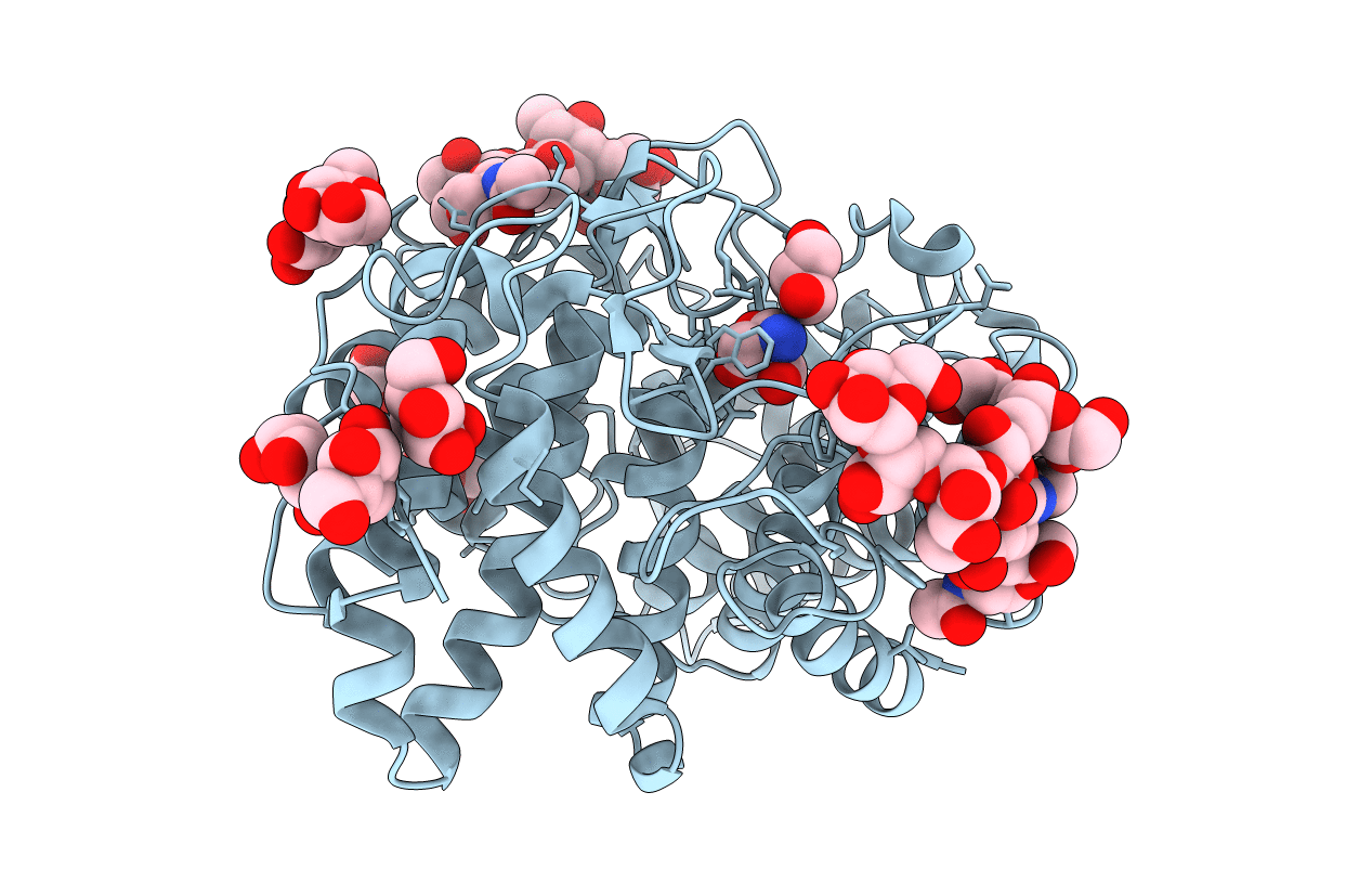
Title:
Catalytic domain of glucoamylase from aspergillus niger complexed with tris and glycerol
Biological Source:
Source Organism(s):
Aspergillus Niger (Taxon ID: 5061)
Method Details:
Experimental Method:
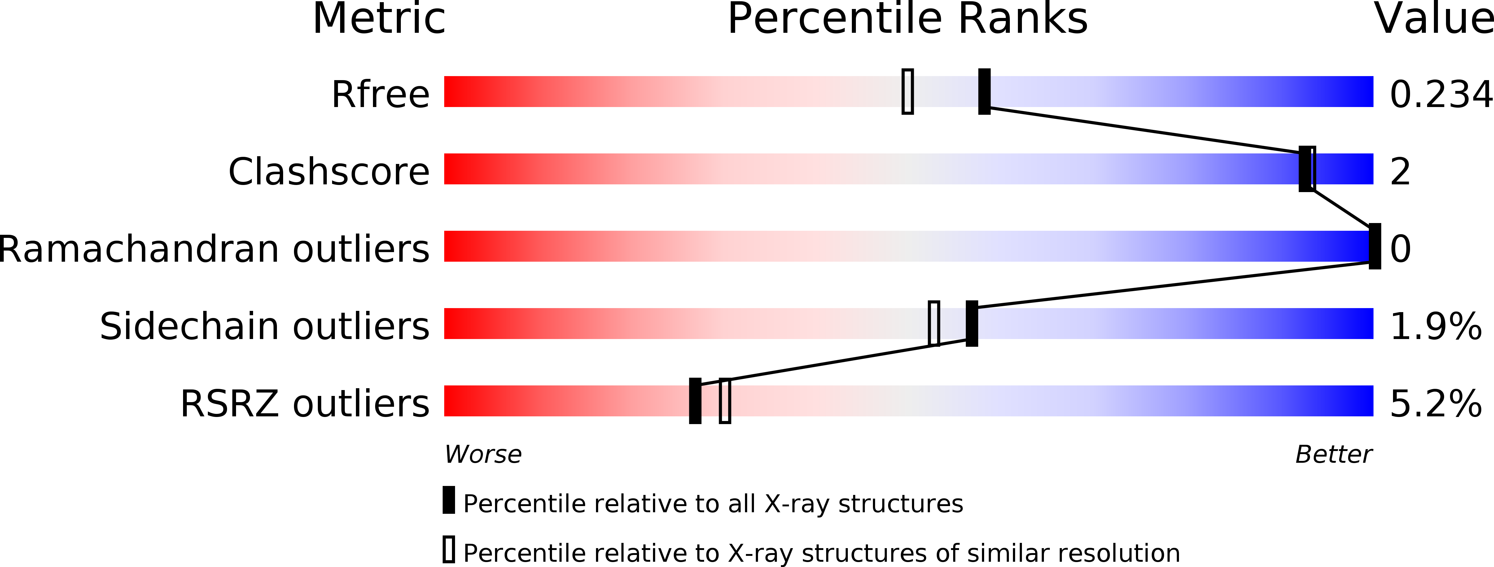
Resolution:
1.90 Å
R-Value Free:
0.22
R-Value Work:
0.18
R-Value Observed:
0.18
Space Group:
P 21 21 21