Deposition Date
2008-09-29
Release Date
2008-11-11
Last Version Date
2024-10-09
Entry Detail
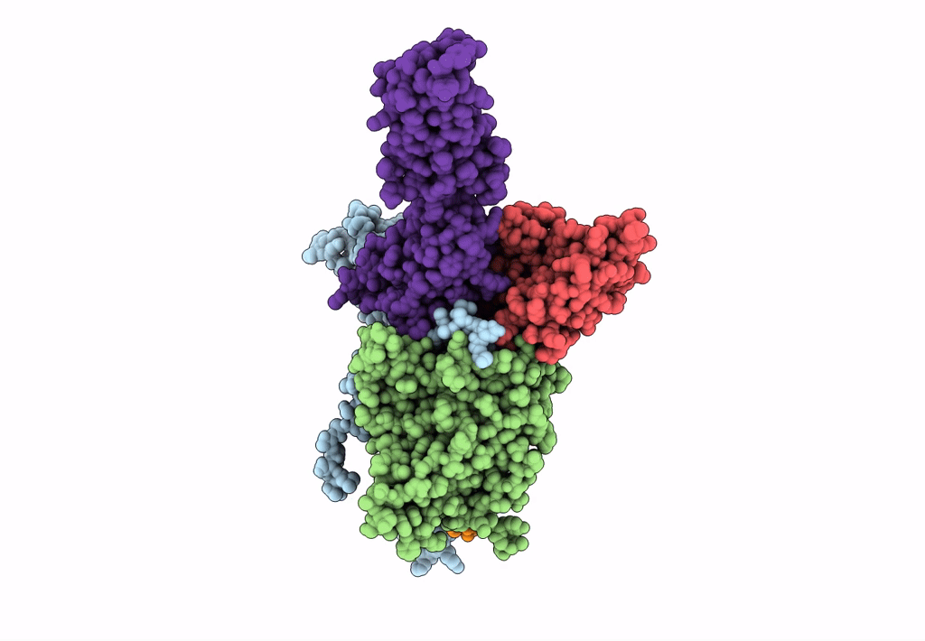
PDB ID:
3EPC
Keywords:
Title:
CryoEM structure of poliovirus receptor bound to poliovirus type 1
Biological Source:
Source Organism(s):
Homo sapiens (Taxon ID: 9606)
Human poliovirus 1 Mahoney (Taxon ID: 12081)
Human poliovirus 1 Mahoney (Taxon ID: 12081)
Expression System(s):
Method Details:
Experimental Method:
Resolution:
8.00 Å
Aggregation State:
PARTICLE
Reconstruction Method:
SINGLE PARTICLE