Deposition Date
2008-09-26
Release Date
2008-10-07
Last Version Date
2024-02-21
Entry Detail
PDB ID:
3EO3
Keywords:
Title:
Crystal structure of the N-acetylmannosamine kinase domain of human GNE protein
Biological Source:
Source Organism(s):
Homo sapiens (Taxon ID: 9606)
Expression System(s):
Method Details:
Experimental Method:
Resolution:
2.84 Å
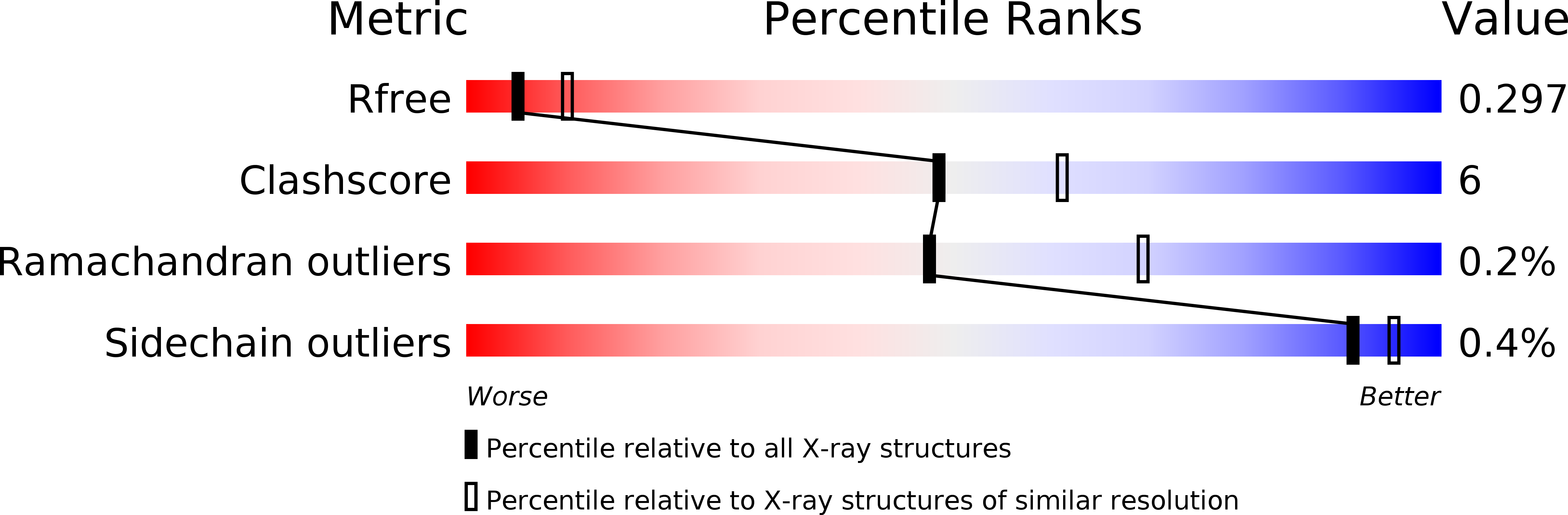
R-Value Free:
0.24
R-Value Work:
0.20
R-Value Observed:
0.20
Space Group:
P 31 2 1