3EMW image
Deposition Date 2008-09-25
Release Date 2008-10-28
Last Version Date 2023-11-01
Entry Detail
PDB ID:
3EMW
Keywords:
Title:
Crystal Structure of human splA/ryanodine receptor domain and SOCS box containing 2 (SPSB2) in complex with a 20-residue VASA peptide
Biological Source:
Source Organism:
Homo sapiens (Taxon ID: 9606)
Host Organism:
Method Details:
Experimental Method:
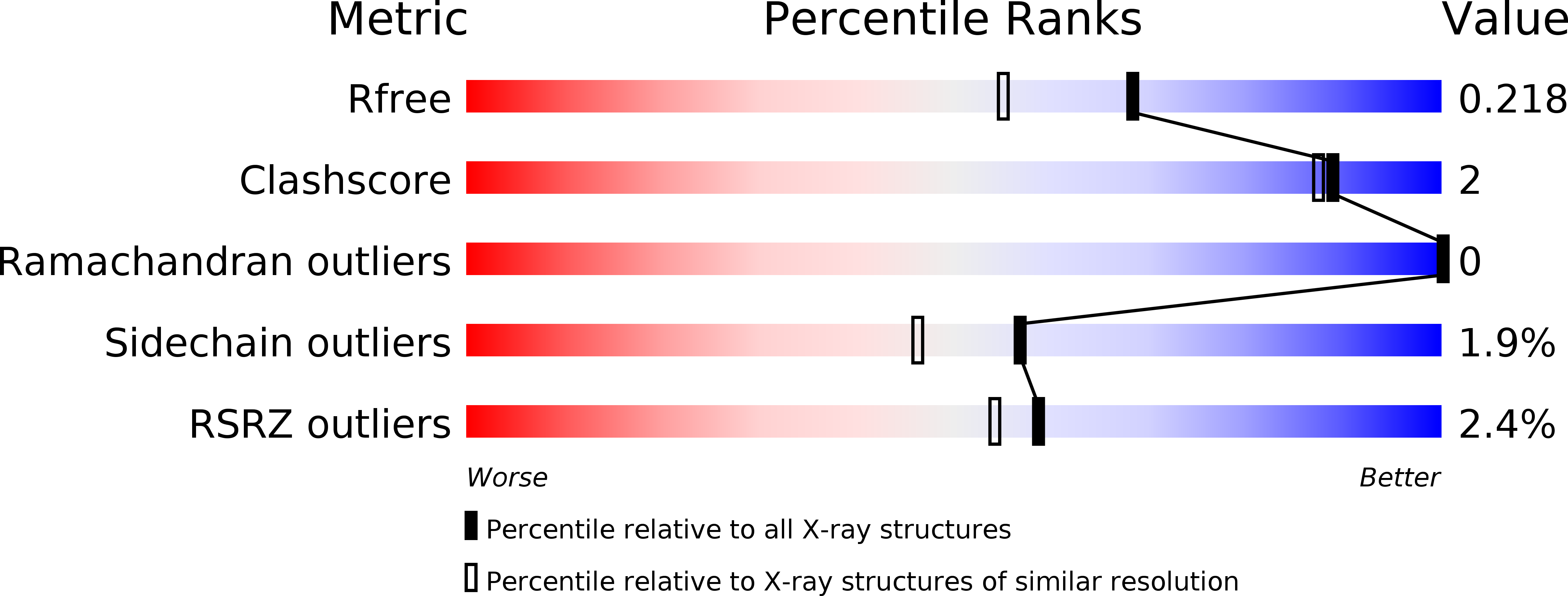
Resolution:
1.80 Å
R-Value Free:
0.21
R-Value Work:
0.17
R-Value Observed:
0.17
Space Group:
P 21 21 21
Macromolecular Entities
Structures with similar UniProt ID
Protein Blast
Polymer Type:polypeptide(L)
Molecule:SPRY domain-containing SOCS box protein 2
Gene (Uniprot):SPSB2
Chain IDs:A
Chain Length:217
Number of Molecules:1
Biological Source:Homo sapiens
Structures with similar UniProt ID
Protein Blast
Polymer Type:polypeptide(L)
Molecule:Peptide (VASA)
Chain IDs:B
Chain Length:20
Number of Molecules:1
Biological Source:
Ligand Molecules
Primary Citation
Structural basis for Par-4 recognition by the SPRY domain- and SOCS box-containing proteins SPSB1, SPSB2, and SPSB4.
J.Mol.Biol. 401 389 402 (2010)
PMID: 20561531 DOI: 10.1016/j.jmb.2010.06.017

Abstact

The mammalian SPRY domain- and SOCS box-containing proteins, SPSB1 to SPSB4, belong to the SOCS box family of E3 ubiquitin ligases. Substrate recognition sites for the SPRY domain are identified only for human Par-4 (ELNNNL) and for the Drosophila orthologue GUSTAVUS binding to the DEAD-box RNA helicase VASA (DINNNN). To further investigate this consensus motif, we determined the crystal structures of SPSB1, SPSB2, and SPSB4, as well as their binding modes and affinities for both Par-4 and VASA. Mutation of each of the three Asn residues in Par-4 abrogated binding to all three SPSB proteins, while changing EL to DI enhanced binding. By comparison to SPSB1 and SPSB4, the more divergent protein SPSB2 showed only weak binding to Par-4 and was hypersensitive to DI substitution. Par-4((59-77)) binding perturbed NMR resonances from a number of SPSB2 residues flanking the ELNNN binding site, including loop D, which binds the EL/DI sequence. Although interactions with the consensus peptide motif were conserved in all structures, flanking sites in SPSB2 were identified as sites of structural change. These structural changes limit high-affinity interactions for SPSB2 to aspartate-containing sequences, whereas SPSB1 and SPSB4 bind strongly to both Par-4 and VASA peptides.

Legend

Protein

Chemical

Disease

Primary Citation of related structures