3ELA image
Entry Detail
PDB ID:
3ELA
Title:
Crystal structure of active site inhibited coagulation factor VIIA mutant in complex with soluble tissue factor
Biological Source:
Source Organism:
Host Organism:
PDB Version:
Deposition Date:
2008-09-21
Release Date:
2008-11-04
Method Details:
Experimental Method:
Resolution:
2.20 Å
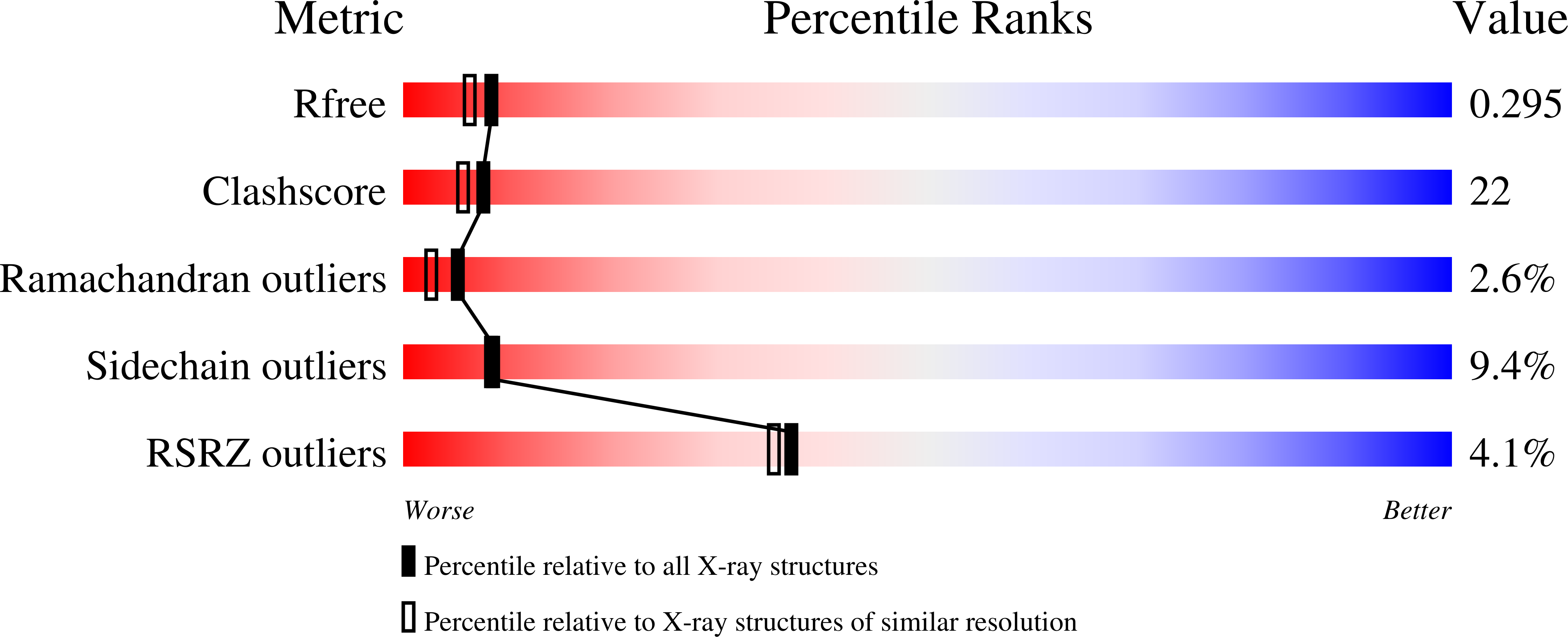
R-Value Free:
0.29
R-Value Work:
0.23
R-Value Observed:
0.23
Space Group:
P 1 21 1
Macromolecular Entities
Polymer Type:polypeptide(L)
Description:Coagulation factor VIIA heavy chain
Mutations:V158D,E296V,M298Q
Chain IDs:B (auth: H)
Chain Length:254
Number of Molecules:1
Biological Source:Homo sapiens
Polymer Type:polypeptide(L)
Description:Coagulation factor VIIA light chain
Chain IDs:A (auth: L)
Chain Length:152
Number of Molecules:1
Biological Source:Homo sapiens
Polymer Type:polypeptide(L)
Description:Tissue factor
Chain IDs:C (auth: T)
Chain Length:209
Number of Molecules:1
Biological Source:Homo sapiens
Peptide-like Molecules
PRD_000369
Primary Citation
Mechanism of the Ca2+-induced enhancement of the intrinsic factor VIIa activity
J.Biol.Chem. 283 25863 25870 (2008)
PMID: 18640965 DOI: 10.1074/jbc.M800841200

Abstact

The intrinsic activity of coagulation factor VIIa (FVIIa) is dependent on Ca(2+) binding to a loop (residues 210-220) in the protease domain. Structural analysis revealed that Ca(2+) may enhance the activity by attenuating electrostatic repulsion of Glu(296) and/or by facilitating interactions between the loop and Lys(161) in the N-terminal tail. In support of the first mechanism, the mutations E296V and D212N resulted in similar, about 2-fold, enhancements of the amidolytic activity. Moreover, mutation of the Lys(161)-interactive residue Asp(217) or Asp(219) to Ala reduced the amidolytic activity by 40-50%, whereas the K161A mutation resulted in 80% reduction. Hence one of these Asp residues in the Ca(2+)-binding loop appears to suffice for some residual interaction with Lys(161), whereas the more severe effect upon replacement of Lys(161) is due to abrogation of the interaction with the N-terminal tail. However, Ca(2+) attenuation of the repulsion between Asp(212) and Glu(296) keeps the activity above that of apoFVIIa. Altogether, our data suggest that repulsion involving Asp(212) in the Ca(2+)-binding loop suppresses FVIIa activity and that optimal activity requires a favorable interaction between the Ca(2+)-binding loop and the N-terminal tail. Crystal structures of tissue factor-bound FVIIa(D212N) and FVIIa(V158D/E296V/M298Q) revealed altered hydrogen bond networks, resembling those in factor Xa and thrombin, after introduction of the D212N and E296V mutations plausibly responsible for tethering the N-terminal tail to the activation domain. The charge repulsion between the Ca(2+)-binding loop and the activation domain appeared to be either relieved by charge removal and new hydrogen bonds (D212N) or abolished (E296V). We propose that Ca(2+) stimulates the intrinsic FVIIa activity by a combination of charge neutralization and loop stabilization.

Legend

Protein

Chemical

Disease

Primary Citation of related structures