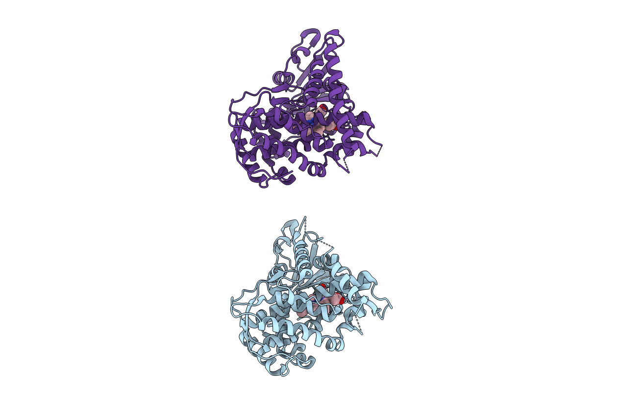
3EKF image
Entry Detail
PDB ID:
3EKF
Keywords:
Title:
Crystal structure of the A264Q heme domain of cytochrome P450 BM3
Biological Source:
Source Organism:
Host Organism:
PDB Version:
Deposition Date:
2008-09-19
Release Date:
2008-12-30
Method Details:
Experimental Method:
Resolution:
2.10 Å
R-Value Free:
0.26
R-Value Work:
0.21
R-Value Observed:
0.21
Space Group:
P 21 21 21
Macromolecular Entities
Polymer Type:polypeptide(L)
Description:Cytochrome P450(BM-3)
Mutations:A264Q
Chain IDs:A, B
Chain Length:470
Number of Molecules:2
Biological Source:Bacillus megaterium
Ligand Molecules
Primary Citation
Novel haem co-ordination variants of flavocytochrome P450BM3.
Biochem.J. 417 65 76 (2009)
PMID: 18721129 DOI: 10.1042/BJ20081133

Abstact

Bacillus megaterium flavocytochrome P450 BM3 is a catalytically self-sufficient fatty acid hydroxylase formed by fusion of soluble NADPH-cytochrome P450 reductase and P450 domains. Selected mutations at residue 264 in the haem (P450) domain of the enzyme lead to novel amino acid sixth (distal) co-ordination ligands to the haem iron. The catalytic, spectroscopic and thermodynamic properties of the A264M, A264Q and A264C variants were determined in both the intact flavocytochromes and haem domains of P450 BM3. Crystal structures of the mutant haem domains demonstrate axial ligation of P450 haem iron by methionine and glutamine ligands trans to the cysteine thiolate, creating novel haem iron ligand sets in the A264M/Q variants. In contrast, the crystal structure of the A264C variant reveals no direct interaction between the introduced cysteine side chain and the haem, although EPR data indicate Cys(264) interactions with haem iron in solution. The A264M haem potential is elevated by comparison with wild-type haem domain, and substrate binding to the A264Q haem domain results in a approximately 360 mV increase in potential. All mutant haem domains occupy the conformation adopted by the substrate-bound form of wild-type BM3, despite the absence of added substrate. The A264M mutant (which has higher dodecanoate affinity than wild-type BM3) co-purifies with a structurally resolved lipid. These data demonstrate that a single mutation at Ala(264) is enough to perturb the conformational equilibrium between substrate-free and substrate-bound P450 BM3, and provide firm structural and spectroscopic data for novel haem iron ligand sets unprecedented in nature.

Legend

Protein

Chemical

Disease

Primary Citation of related structures