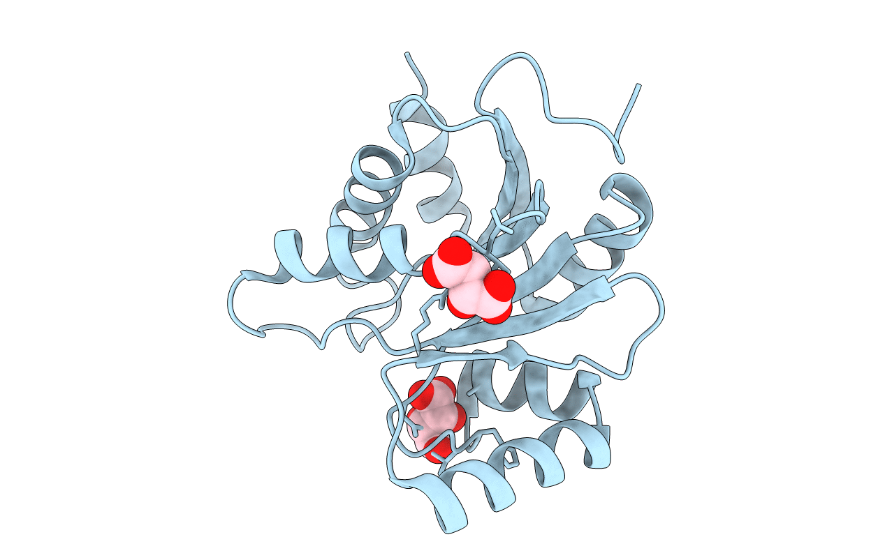
3EKE image
Deposition Date 2008-09-19
Release Date 2008-09-30
Last Version Date 2024-02-21
Entry Detail
PDB ID:
3EKE
Keywords:
Title:
Crystal structure of IBV X-domain at pH 5.6
Biological Source:
Method Details:
Experimental Method:
Resolution:
2.10 Å
R-Value Free:
0.23
R-Value Work:
0.19
R-Value Observed:
0.20
Space Group:
P 32 2 1
Macromolecular Entities
Polymer Type:polypeptide(L)
Molecule:Non-structural protein 3
Gene (Uniprot):rep
Chain IDs:A
Chain Length:176
Number of Molecules:1
Biological Source:Avian infectious bronchitis virus (strain Beaudette)
Ligand Molecules
Primary Citation
Crystal structures of the X-domains of a Group-1 and a Group-3 coronavirus reveal that ADP-ribose-binding may not be a conserved property.
Protein Sci. 18 6 16 (2009)
PMID: 19177346 DOI: 10.1002/pro.15

Abstact

The polyproteins of coronaviruses are cleaved by viral proteases into at least 15 nonstructural proteins (Nsps). Consisting of five domains, Nsp3 is the largest of these (180-210 kDa). Among these domains, the so-called X-domain is believed to act as ADP-ribose-1''-phosphate phosphatase or to bind poly(ADP-ribose). However, here we show that the X-domain of Infectious Bronchitis Virus (strain Beaudette), a Group-3 coronavirus, fails to bind ADP-ribose. This is explained on the basis of the crystal structure of the protein, determined at two different pH values. For comparison, we also describe the crystal structure of the homologous X-domain from Human Coronavirus 229E, a Group-1 coronavirus, which does bind ADP-ribose.

Legend

Protein

Chemical

Disease

Primary Citation of related structures