3EK9 image
Deposition Date 2008-09-19
Release Date 2009-02-24
Last Version Date 2023-08-30
Entry Detail
PDB ID:
3EK9
Keywords:
Title:
SPRY Domain-containing SOCS Box Protein 2: Crystal Structure and Residues Critical for Protein Binding
Biological Source:
Source Organism:
Mus musculus (Taxon ID: 10090)
Host Organism:
Method Details:
Experimental Method:
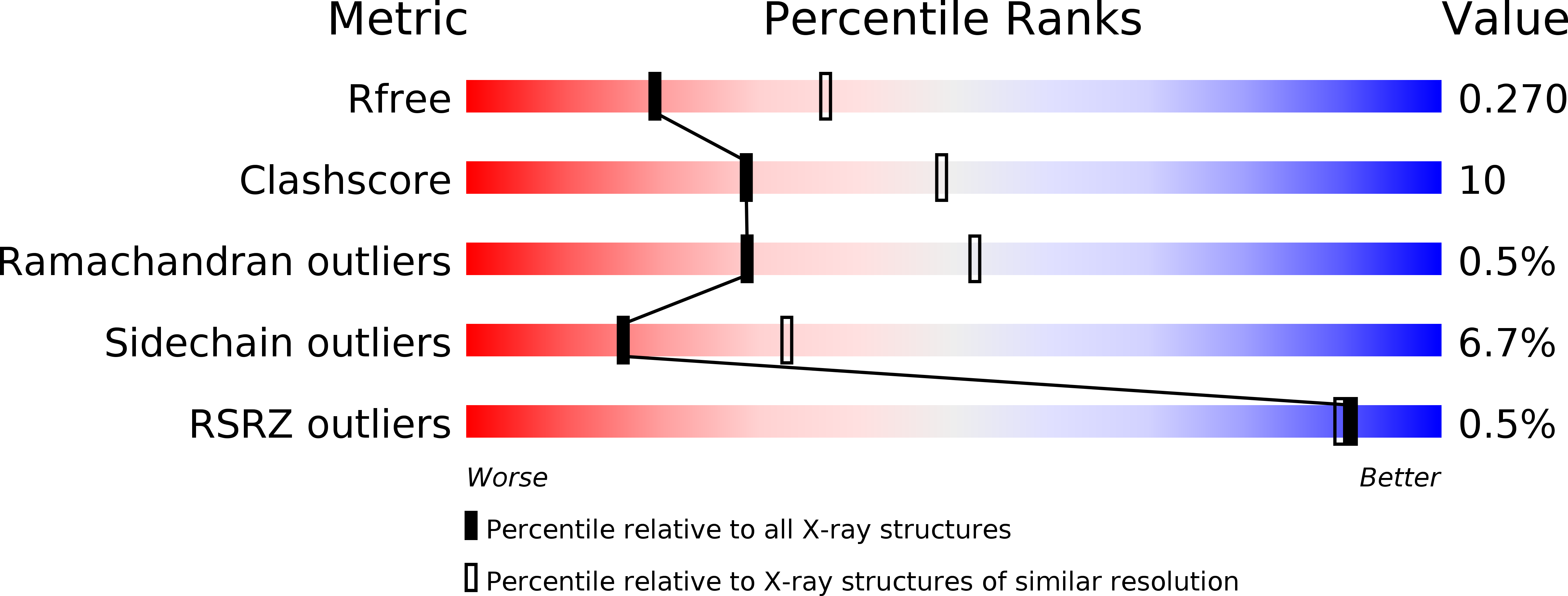
Resolution:
2.60 Å
R-Value Free:
0.27
R-Value Work:
0.16
R-Value Observed:
0.17
Space Group:
P 21 21 21
Macromolecular Entities
Polymer Type:polypeptide(L)
Molecule:SPRY domain-containing SOCS box protein 2
Gene (Uniprot):Spsb2
Chain IDs:A
Chain Length:213
Number of Molecules:1
Biological Source:Mus musculus
Ligand Molecules
Primary Citation
SPRY domain-containing SOCS box protein 2: crystal structure and residues critical for protein binding.
J.Mol.Biol. 386 662 674 (2009)
PMID: 19154741 DOI: 10.1016/j.jmb.2008.12.078

Abstact

The four mammalian SPRY (a sequence repeat in dual-specificity kinase splA and ryanodine receptors) domain-containing suppressor of cytokine signalling (SOCS) box proteins (SSB-1 to -4) are characterised by a C-terminal SOCS box and a central SPRY domain. The latter is a protein interaction module found in over 1600 proteins, with more than 70 encoded in the human genome. Here we report the crystal structure of the SPRY domain of murine SSB-2 and compare it with the SSB-2 solution structure and crystal structures of other B30.2/SPRY domain-containing family proteins. The structure is a bent beta-sandwich, consisting of two seven-stranded beta-sheets wrapped around a long loop that extends from the centre strands of the inner or concave beta-sheet; it closely matches those of GUSTAVUS and SSB-4. The structure is also similar to those of two recently determined Neuralized homology repeat (NHR) domains (also known as NEUZ domains), with detailed comparisons, suggesting that the NEUZ/NHR domains form a subclass of SPRY domains. The binding site on SSB-2 for the prostate apoptosis response-4 (Par-4) protein has been mapped in finer detail using mutational analyses. Moreover, SSB-1 was shown to have a Par-4 binding surface similar to that identified for SSB-2. Structural perturbations of SSB-2 induced by mutations affecting its interaction with Par-4 and/or c-Met have been characterised by NMR. These comparisons, in conjunction with previously published dynamics data from NMR relaxation studies and coarse-grained dynamics simulation using normal mode analysis, further refine our understanding of the structural basis for protein recognition of SPRY domain-containing proteins.

Legend

Protein

Chemical

Disease

Primary Citation of related structures