3EJJ image
Deposition Date 2008-09-18
Release Date 2008-12-09
Last Version Date 2024-11-20
Entry Detail
PDB ID:
3EJJ
Title:
Structure of M-CSF bound to the first three domains of FMS
Biological Source:
Source Organism:
Mus musculus (Taxon ID: 10090)
Host Organism:
Method Details:
Experimental Method:
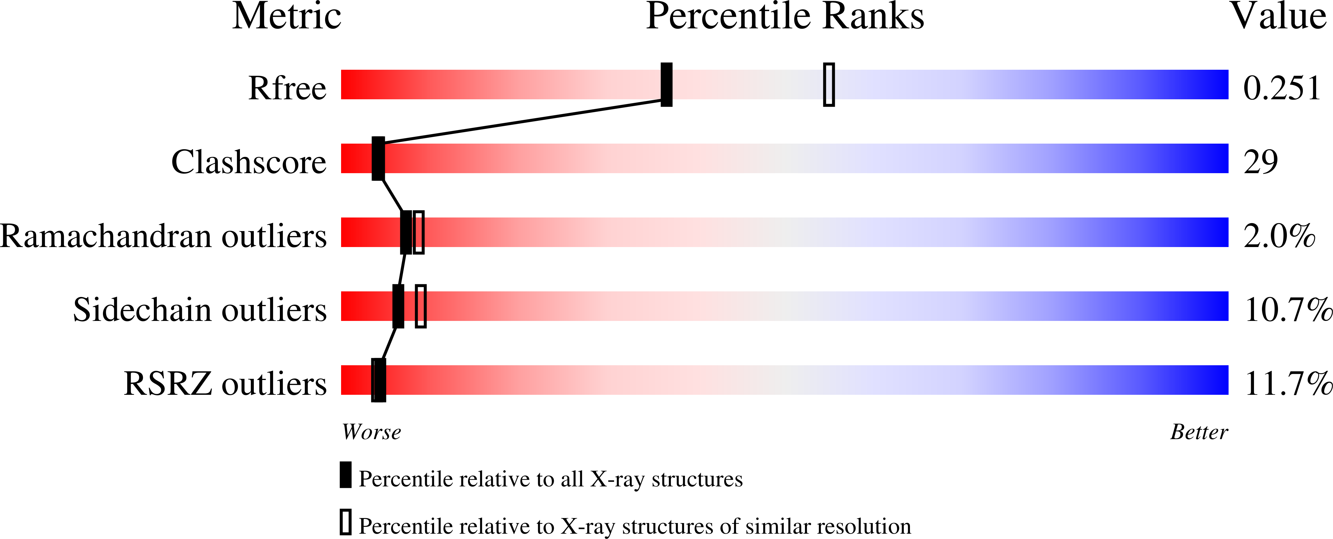
Resolution:
2.40 Å
R-Value Free:
0.26
R-Value Work:
0.23
R-Value Observed:
0.24
Space Group:
H 3 2
Macromolecular Entities
Structures with similar UniProt ID
Protein Blast
Polymer Type:polypeptide(L)
Molecule:Colony stimulating factor-1
Chain IDs:A, B
Chain Length:155
Number of Molecules:2
Biological Source:Mus musculus
Structures with similar UniProt ID
Protein Blast
Polymer Type:polypeptide(L)
Molecule:Macrophage colony-stimulating factor 1 receptor
Gene (Uniprot):Csf1r
Chain IDs:C (auth: X)
Chain Length:289
Number of Molecules:1
Biological Source:Mus musculus
Modified Residue
Compound ID Chain ID Parent Comp ID Details 2D Image
ASN C ASN GLYCOSYLATION SITE
Primary Citation
Structure of macrophage colony stimulating factor bound to FMS: diverse signaling assemblies of class III receptor tyrosine kinases.
Proc.Natl.Acad.Sci.USA 105 18267 18272 (2008)
PMID: 19017797 DOI: 10.1073/pnas.0807762105

Abstact

Macrophage colony stimulating factor (M-CSF), through binding to its receptor FMS, a class III receptor tyrosine kinase (RTK), regulates the development and function of mononuclear phagocytes, and plays important roles in innate immunity, cancer and inflammation. We report a 2.4 A crystal structure of M-CSF bound to the first 3 domains (D1-D3) of FMS. The ligand binding mode of FMS is surprisingly different from KIT, another class III RTK, in which the major ligand-binding domain of FMS, D2, uses the CD and EF loops, but not the beta-sheet on the opposite side of the Ig domain as in KIT, to bind ligand. Calorimetric data indicate that M-CSF cannot dimerize FMS without receptor-receptor interactions mediated by FMS domains D4 and D5. Consistently, the structure contains only 1 FMS-D1-D3 molecule bound to a M-CSF dimer, due to a weak, hydrophilic M-CSF:FMS interface, and probably a conformational change of the M-CSF dimer in which binding to the second site is rendered unfavorable by FMS binding at the first site. The partial, intermediate complex suggests that FMS may be activated in two steps, with the initial engagement step distinct from the subsequent dimerization/activation step. Hence, the formation of signaling class III RTK complexes can be diverse, engaging various modes of ligand recognition and various mechanistic steps for dimerizing and activating receptors.

Legend

Protein

Chemical

Disease

Primary Citation of related structures