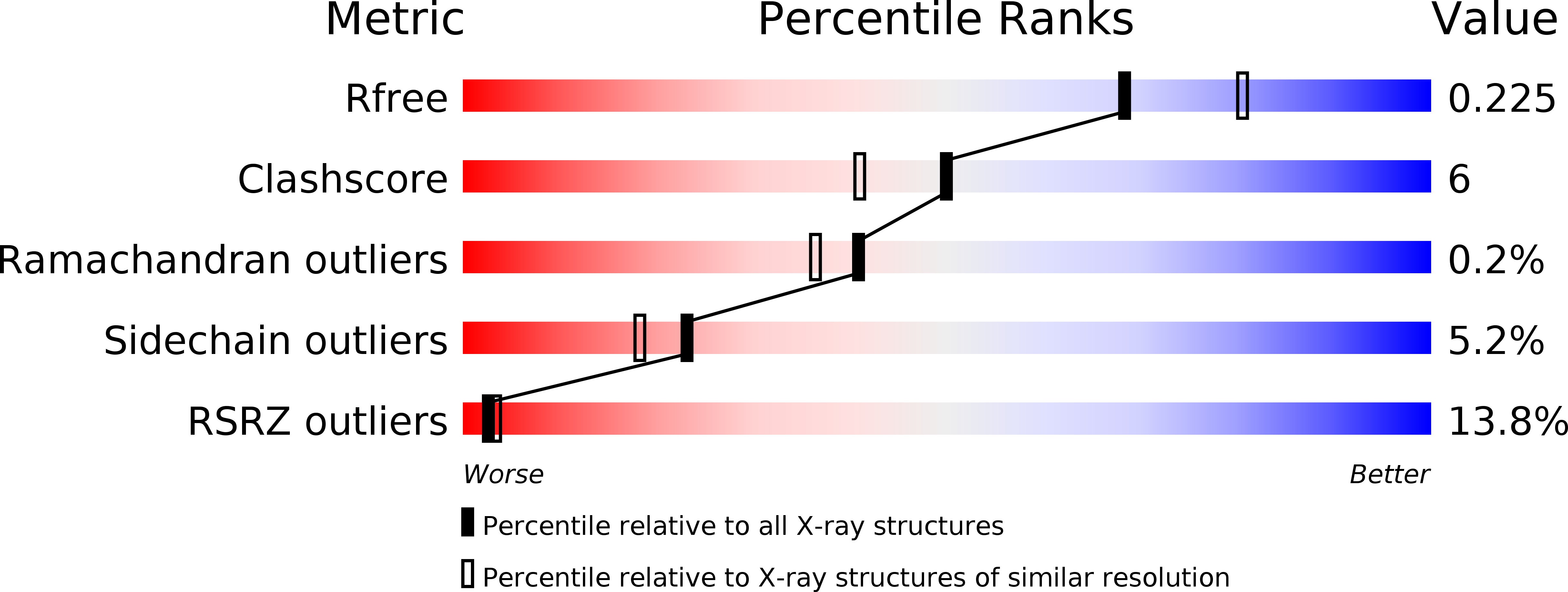
3EIV image
Deposition Date 2008-09-17
Release Date 2009-09-08
Last Version Date 2023-08-30
Entry Detail
PDB ID:
3EIV
Title:
Crystal Structure of Single-stranded DNA-binding protein from Streptomyces coelicolor
Biological Source:
Source Organism:
Method Details:
Experimental Method:
Resolution:
2.14 Å
R-Value Free:
0.25
R-Value Work:
0.23
R-Value Observed:
0.23
Space Group:
I 2 2 2
Macromolecular Entities
Structures with similar UniProt ID
Protein Blast
Polymer Type:polypeptide(L)
Molecule:Single-stranded DNA-binding protein 2
Gene (Uniprot):ssb2
Chain IDs:A, B, C, D
Chain Length:199
Number of Molecules:4
Biological Source:Streptomyces coelicolor
Primary Citation
Structure of the single-stranded DNA-binding protein from Streptomyces coelicolor.
Acta Crystallogr.,Sect.D 65 974 979 (2009)
PMID: 19690375 DOI: 10.1107/S0907444909023634

Abstact

The crystal structure of the single-stranded DNA-binding protein (SSB) from Streptomyces coelicolor, a filamentous soil bacterium with a complex life cycle and a linear chromosome, has been solved and refined at 2.1 A resolution. The three-dimensional structure shows a common conserved central OB-fold that is found in all structurally determined SSB proteins. However, it shows variations in quaternary structure that have previously only been found in mycobacterial SSBs. The strand involved in the clamp mechanism characteristic of this type of quaternary structure leads to higher stability of the homotetramer. To the best of our knowledge, this is the first X-ray structure of an SSB protein from a member of the genus Streptomyces and it was predicted to be the most stable of the structurally characterized bacterial or human mitochondrial SSBs.

Legend

Protein

Chemical

Disease

Primary Citation of related structures