3EIP image
Deposition Date 1999-03-29
Release Date 1999-11-10
Last Version Date 2023-12-27
Entry Detail
PDB ID:
3EIP
Keywords:
Title:
CRYSTAL STRUCTURE OF COLICIN E3 IMMUNITY PROTEIN: AN INHIBITOR TO A RIBOSOME-INACTIVATING RNASE
Biological Source:
Method Details:
Experimental Method:
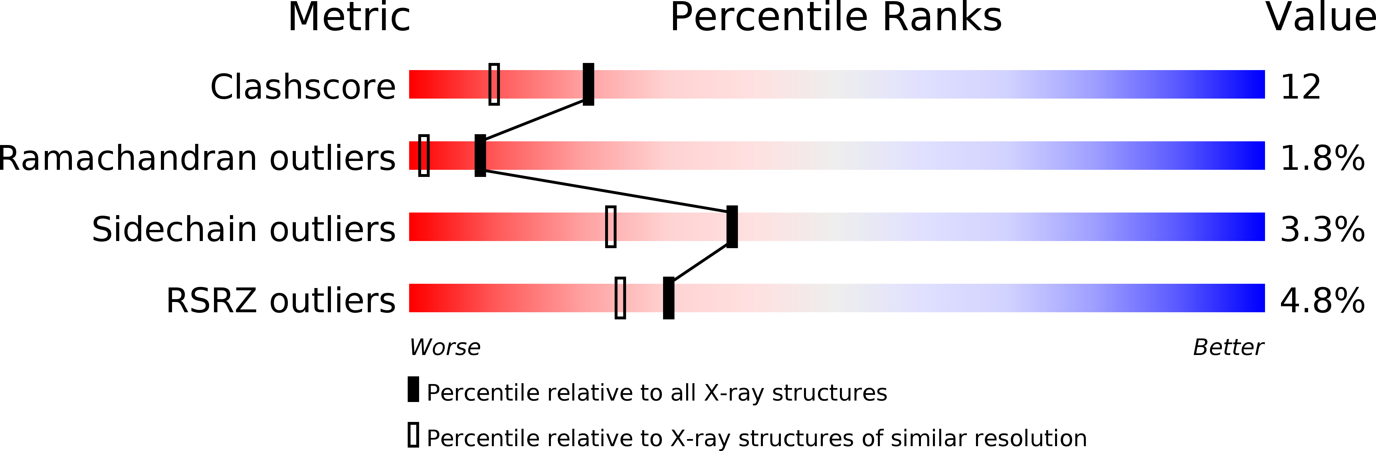
Resolution:
1.80 Å
R-Value Free:
0.26
R-Value Work:
0.21
Space Group:
C 1 2 1
Macromolecular Entities
Polymer Type:polypeptide(L)
Molecule:PROTEIN (COLICIN E3 IMMUNITY PROTEIN)
Gene (Uniprot):imm
Chain IDs:A, B
Chain Length:84
Number of Molecules:2
Biological Source:Escherichia coli str. K12 substr.
Ligand Molecules
Primary Citation
Crystal structure of colicin E3 immunity protein: an inhibitor of a ribosome-inactivating RNase.
Structure Fold.Des. 7 1365 1372 (1999)
PMID: 10574790 DOI: 10.1016/S0969-2126(00)80026-X

Abstact

BACKGROUND: Colicins are antibiotic-like proteins of Escherichia coli that kill related strains. Colicin E3 acts as an RNase that specifically cleaves 16S rRNA, thereby inactivating the ribosomes in the infected cell. The producing organism is protected against colicin E3 by a specific inhibitor, the immunity protein Im3, which forms a tight 1:1 complex with colicin E3 and renders it inactive. Crystallographic studies on colicin E3 and Im3 have been undertaken to unravel the structural basis for the ribonucleolytic activity and its inhibition. RESULTS: The crystal structure of Im3 has been determined to a resolution of 1.8 A. The structure consists of a four-standard antiparallel beta sheet flanked by three alpha helices on one side of the sheet. Thr7, Phe9, Phe16 and Phe74 form a hydrophobic cluster on the surface of the protein in the vicinity of Cys47. This cluster is part of a putative binding pocket which also includes nine polar residues. CONCLUSIONS: The putative binding pocket of Im3 is the probable site of interaction with colicin E3. The six acidic residues in the pocket may interact with some of the numerous basic residues of colicin E3. The involvement of hydrophobic moieties in the binding is consistent with the observation that the tight complex can only be dissociated by denaturation. The structure of Im3 resembles those of certain nucleic acid binding proteins, in particular domain II of topoisomerase I and RNA-binding proteins that contain the ribonucleoprotein (RNP) sequence motif. This observation suggests that Im3 has a nucleic acid binding function in addition to binding colicin E3.

Legend

Protein

Chemical

Disease

Primary Citation of related structures