3EHB image
Deposition Date 2008-09-12
Release Date 2008-09-30
Last Version Date 2025-04-30
Entry Detail
PDB ID:
3EHB
Title:
A D-Pathway Mutation Decouples the Paracoccus Denitrificans Cytochrome c Oxidase by Altering the side chain orientation of a distant, conserved Glutamate
Biological Source:
Source Organism:
Host Organism:
Method Details:
Experimental Method:
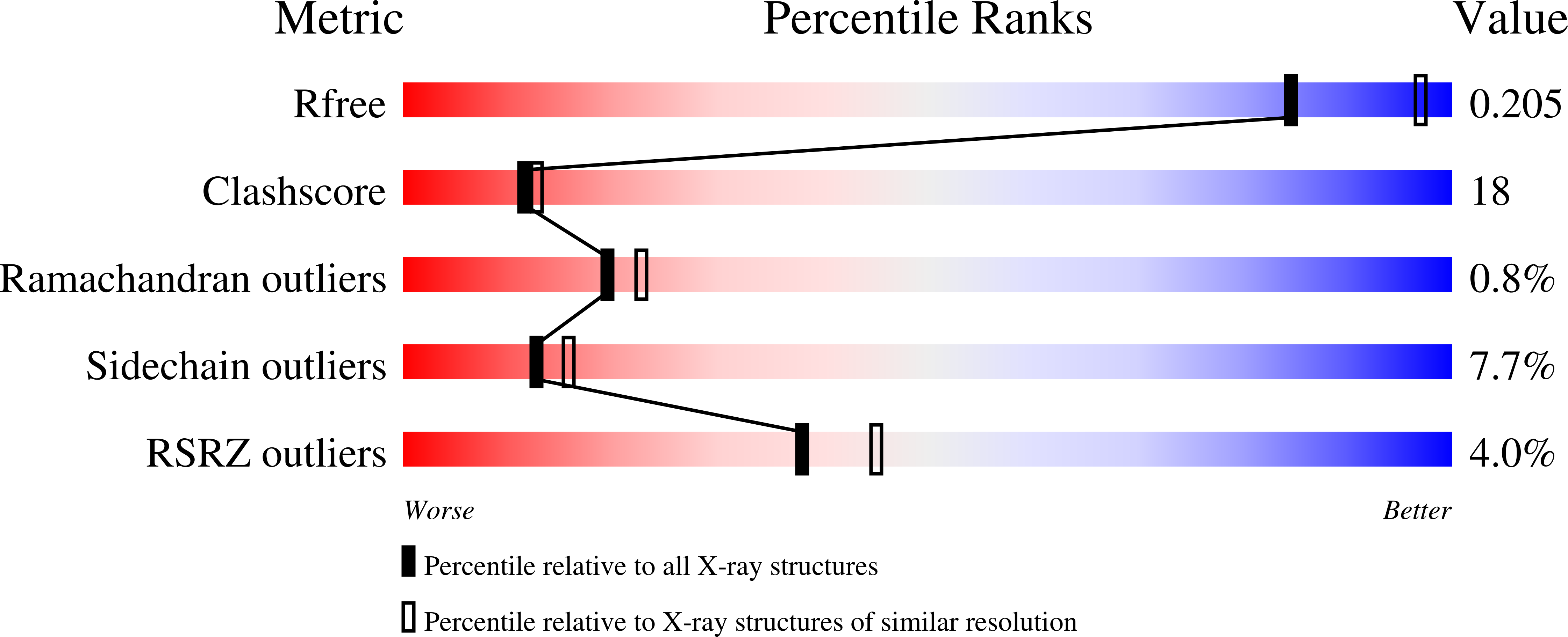
Resolution:
2.32 Å
R-Value Free:
0.24
R-Value Work:
0.20
R-Value Observed:
0.20
Space Group:
P 21 21 21
Macromolecular Entities
Polymer Type:polypeptide(L)
Molecule:Cytochrome c oxidase subunit 1-beta
Gene (Uniprot):ctaDII
Mutations:N131D
Chain IDs:A
Chain Length:558
Number of Molecules:1
Biological Source:Paracoccus denitrificans
Polymer Type:polypeptide(L)
Molecule:Cytochrome c oxidase subunit 2
Gene (Uniprot):ctaC
Chain IDs:B
Chain Length:298
Number of Molecules:1
Biological Source:Paracoccus denitrificans
Polymer Type:polypeptide(L)
Molecule:FV fragment Chain H
Chain IDs:C
Chain Length:127
Number of Molecules:1
Biological Source:Mus musculus
Polymer Type:polypeptide(L)
Molecule:FV fragment Chain L
Chain IDs:D
Chain Length:120
Number of Molecules:1
Biological Source:Mus musculus
Primary Citation
A d-pathway mutation decouples the paracoccusdenitrificans cytochrome C oxidase by altering the side-chain orientation of a distant conserved glutamate.
J.Mol.Biol. 384 865 877 (2008)
PMID: 18930738 DOI: 10.1016/j.jmb.2008.09.074

Abstact

Asparagine 131, located near the cytoplasmic entrance of the D-pathway in subunit I of the Paracoccus denitrificans aa(3) cytochrome c oxidase, is a residue crucial for proton pumping. When replaced by an aspartate, the mutant enzyme is completely decoupled: while retaining full cytochrome c oxidation activity, it does not pump protons. The same phenotype is observed for two other substitutions at this position (N131E and N131C), whereas a conservative replacement by glutamine affects both activities of the enzyme. The N131D variant oxidase was crystallized and its structure was solved to 2.32-A resolution, revealing no significant overall change in the protein structure when compared with the wild type (WT), except for an alternative orientation of the E278 side chain in addition to its WT conformation. Moreover, remarkable differences in the crystallographically resolved chain of water molecules in the D-pathway are found for the variant: four water molecules that are observed in the water chain between N131 and E278 in the WT structure are not visible in the variant, indicating a higher mobility of these water molecules. Electrochemically induced Fourier transform infrared difference spectra of decoupled mutants confirm that the protonation state of E278 is unaltered by these mutations but indicate a distinct perturbation in the hydrogen-bonding environment of this residue. Furthermore, they suggest that the carboxylate side chain of the N131D mutant is deprotonated. These findings are discussed in terms of their mechanistic implications for proton routing through the D-pathway of cytochrome c oxidase.

Legend

Protein

Chemical

Disease

Primary Citation of related structures