Deposition Date
2008-09-11
Release Date
2009-04-28
Last Version Date
2023-08-30
Entry Detail
PDB ID:
3EHA
Keywords:
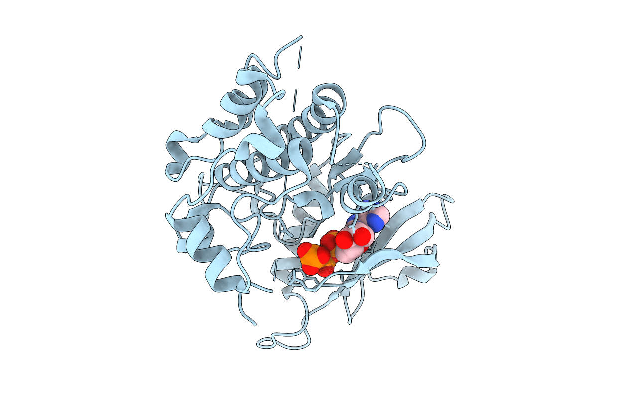
Title:
Crystal structure of death associated protein kinase complexed with AMPPNP
Biological Source:
Source Organism(s):
Homo sapiens (Taxon ID: 9606)
Expression System(s):
Method Details:
Experimental Method:
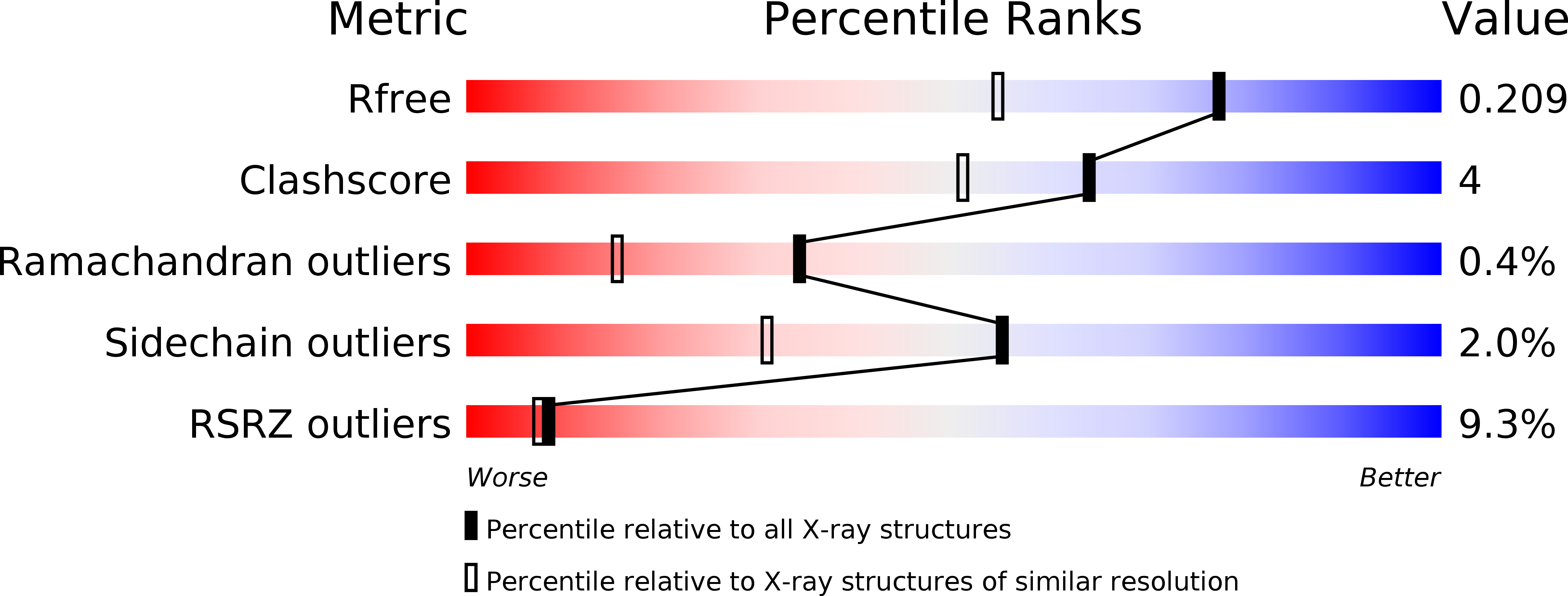
Resolution:
1.60 Å
R-Value Free:
0.21
R-Value Work:
0.17
R-Value Observed:
0.17
Space Group:
P 21 21 21