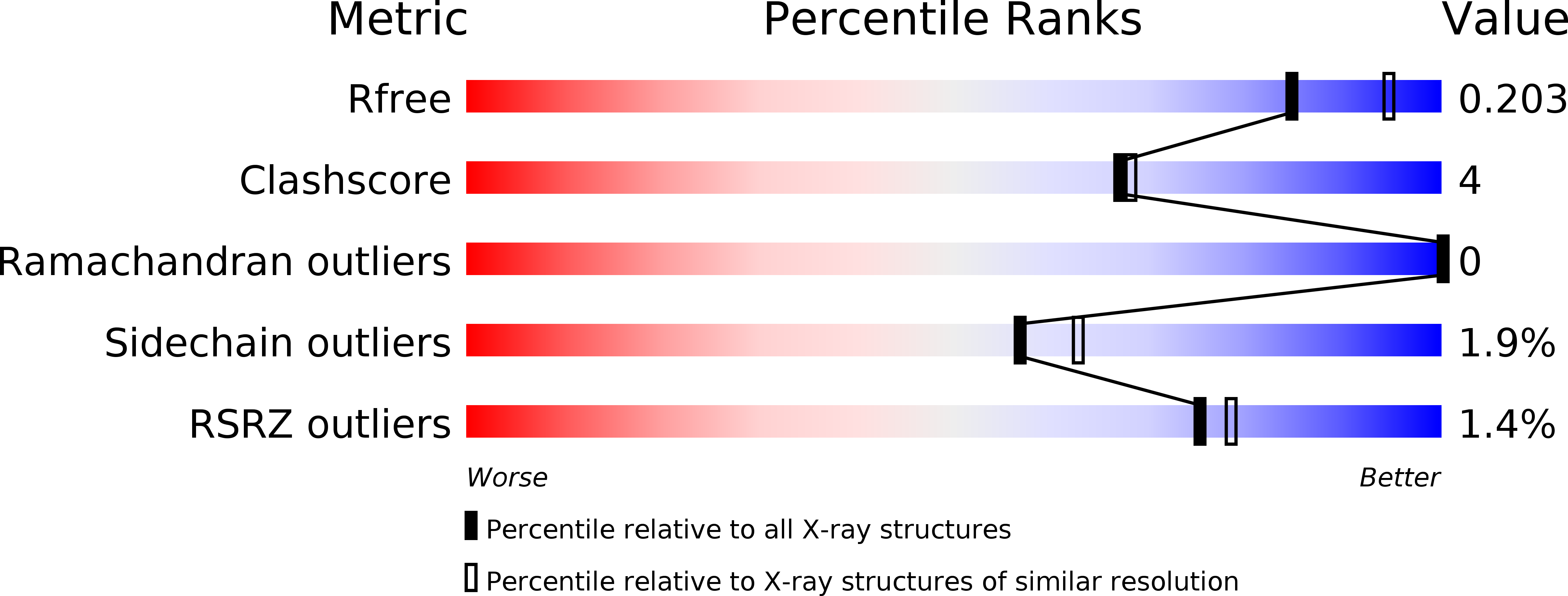
3EGM image
Deposition Date 2008-09-11
Release Date 2009-07-28
Last Version Date 2024-03-20
Entry Detail
PDB ID:
3EGM
Keywords:
Title:
Structural basis of iron transport gating in Helicobacter pylori ferritin
Biological Source:
Source Organism:
Host Organism:
Method Details:
Experimental Method:
Resolution:
2.10 Å
R-Value Free:
0.20
R-Value Work:
0.16
R-Value Observed:
0.16
Space Group:
I 4
Macromolecular Entities
Structures with similar UniProt ID
Protein Blast
Polymer Type:polypeptide(L)
Molecule:Ferritin
Gene (Uniprot):ftnA
Chain IDs:A, B, C, D, E, F
Chain Length:181
Number of Molecules:6
Biological Source:Helicobacter pylori
Primary Citation
The crystal structure of ferritin from Helicobacter pylori reveals unusual conformational changes for iron uptake.
J.Mol.Biol. 390 83 98 (2009)
PMID: 19427319 DOI: 10.1016/j.jmb.2009.04.078

Abstact

The crystal structure of recombinant ferritin from Helicobacter pylori has been determined in its apo, low-iron-bound, intermediate, and high-iron-bound states. Similar to other members of the ferritin family, the bacterial ferritin assembles as a spherical protein shell of 24 subunits, each of which folds into a four-alpha-helix bundle. Significant conformational changes were observed at the BC loop and the entrance of the 4-fold symmetry channel in the intermediate and high-iron-bound states, whereas no change was found in the apo and low-iron-bound states. The imidazole rings of His149 at the channel entrance undergo conformational changes that bear resemblance to heme configuration and are directly coupled to axial translocation of Fe ions through the 4-fold channel. Our results provide the first structural evidence of the translocation of Fe ions through the 4-fold channel in prokaryotes and the transition from a protein-dominated process to a mineral-surface-dominated process during biomineralization.

Legend

Protein

Chemical

Disease

Primary Citation of related structures