Deposition Date
2008-09-10
Release Date
2008-10-14
Last Version Date
2023-11-01
Entry Detail
PDB ID:
3EG5
Keywords:
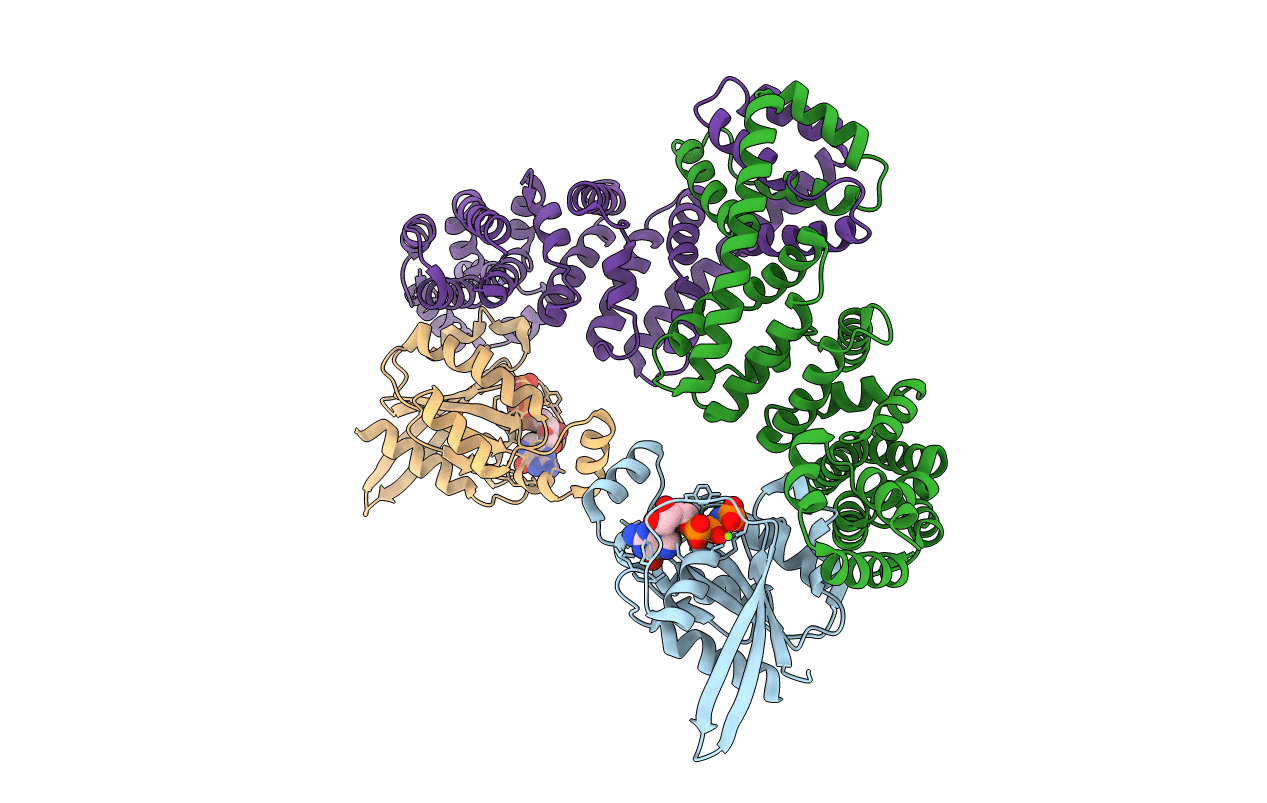
Title:
Crystal structure of MDIA1-TSH GBD-FH3 in complex with CDC42-GMPPNP
Biological Source:
Source Organism(s):
Mus musculus (Taxon ID: 10090)
Expression System(s):
Method Details:
Experimental Method:
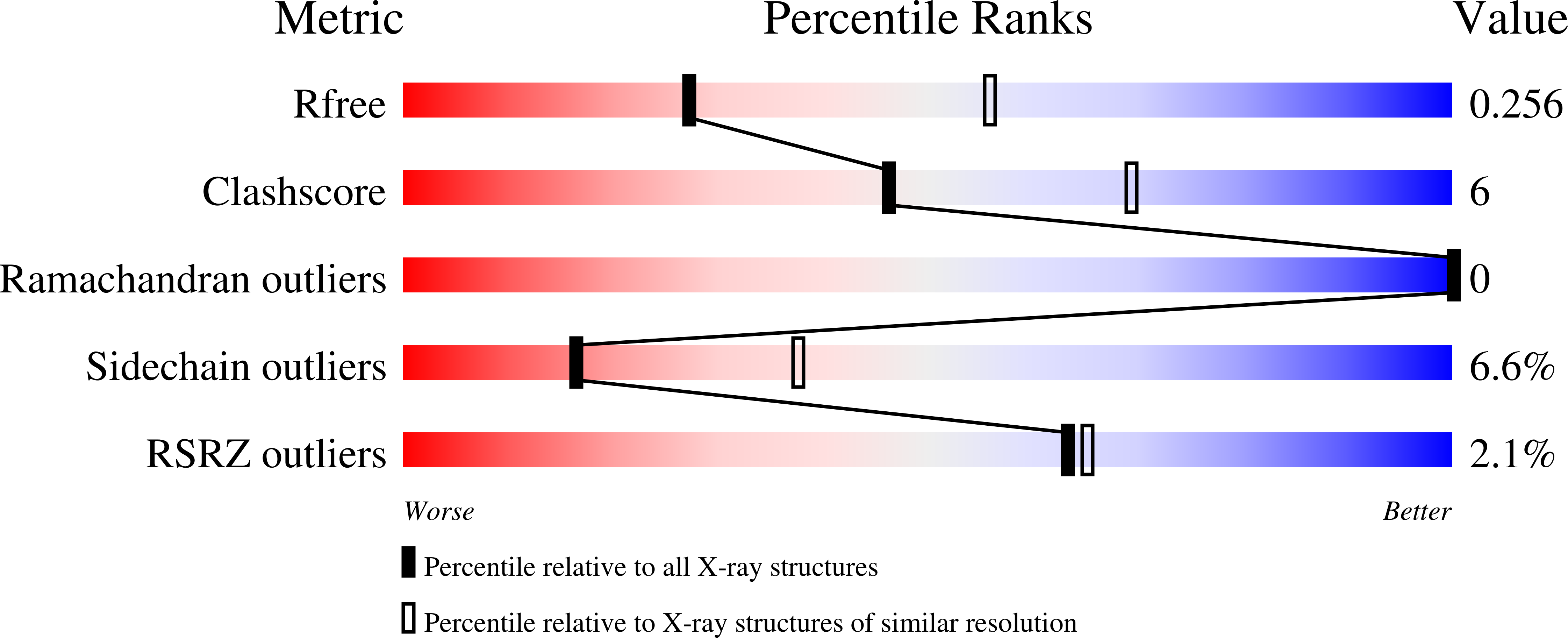
Resolution:
2.70 Å
R-Value Free:
0.24
R-Value Work:
0.20
R-Value Observed:
0.20
Space Group:
P 32