3EFZ image
Deposition Date 2008-09-10
Release Date 2008-09-23
Last Version Date 2024-10-30
Entry Detail
PDB ID:
3EFZ
Title:
Crystal Structure of a 14-3-3 protein from cryptosporidium parvum (cgd1_2980)
Biological Source:
Source Organism:
Host Organism:
Method Details:
Experimental Method:
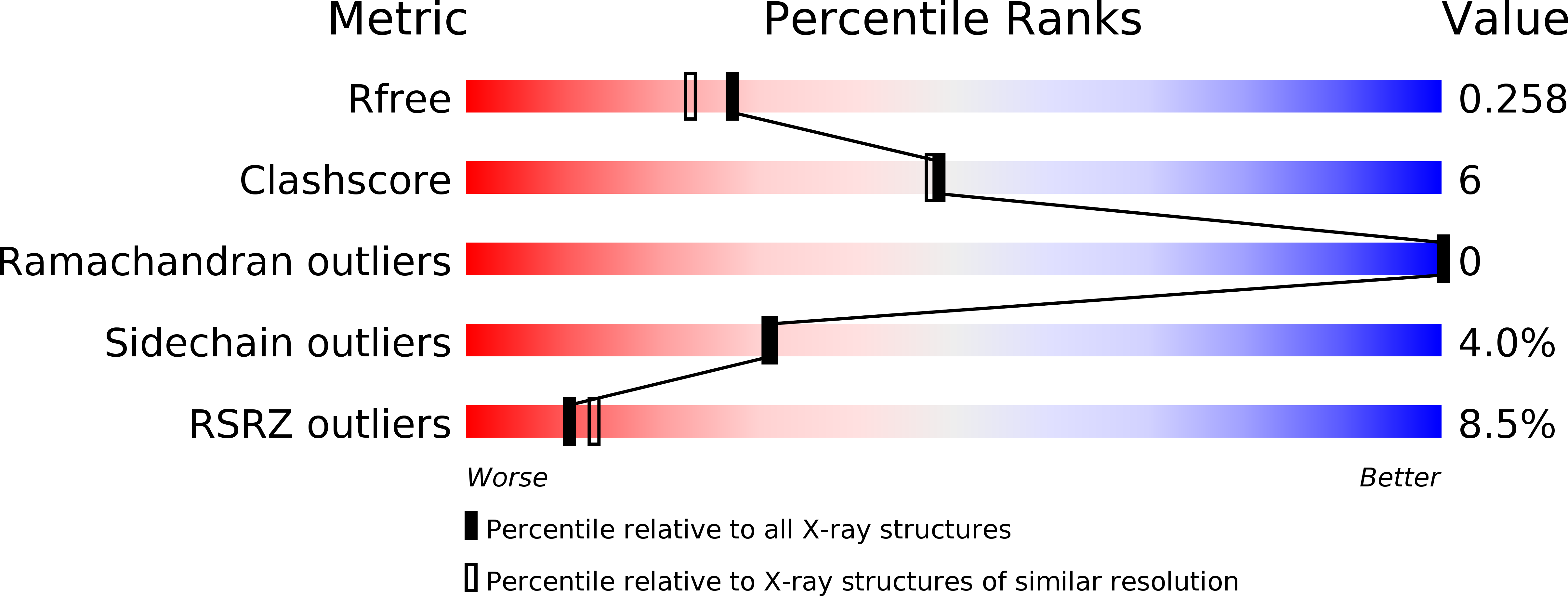
Resolution:
2.08 Å
R-Value Free:
0.26
R-Value Work:
0.22
R-Value Observed:
0.22
Space Group:
P 1 21 1
Macromolecular Entities
Structures with similar UniProt ID
Protein Blast
Polymer Type:polypeptide(L)
Molecule:14-3-3 protein
Gene (Uniprot):cgd1_2980
Chain IDs:A, B, C, D
Chain Length:268
Number of Molecules:4
Biological Source:Cryptosporidium parvum
Modified Residue
Compound ID Chain ID Parent Comp ID Details 2D Image
SEP A SER PHOSPHOSERINE
Ligand Molecules
Primary Citation
Characterization of 14-3-3 proteins from Cryptosporidium parvum.
Plos One 6 e14827 e14827 (2011)
PMID: 21853016 DOI: 10.1371/journal.pone.0014827

Abstact

UNLABELLED The parasite Cryptosporidium parvum has three 14-3-3 proteins: Cp14ε, Cp14a and Cp14b, with only Cp14ε similar to human 14-3-3 proteins in sequence, peptide-binding properties and structure. Structurally, Cp14a features the classical 14-3-3 dimer but with a uniquely wide pocket and a disoriented RRY triad potentially incapable of binding phosphopeptides. The Cp14b protein deviates from the norm significantly: (i) In one subunit, the phosphorylated C-terminal tail is bound in the binding groove like a phosphopeptide. This supports our binding study indicating this protein was stabilized by a peptide mimicking its last six residues. (ii) The other subunit has eight helices instead of nine, with αA and αB forming a single helix and occluding the peptide-binding cleft. (iii) The protein forms a degenerate dimer with the two binding grooves divided and facing opposite directions. These features conspire to block and disrupt the bicameral substrate-binding pocket, suggesting a possible tripartite auto-regulation mechanism that has not been observed previously. ENHANCED VERSION This article can also be viewed as an enhanced version in which the text of the article is integrated with interactive 3D representations and animated transitions. Please note that a web plugin is required to access this enhanced functionality. Instructions for the installation and use of the web plugin are available in Text S1.

Legend

Protein

Chemical

Disease

Primary Citation of related structures