3EFF image
Entry Detail
PDB ID:
3EFF
Title:
The Crystal Structure of Full-Length KcsA in its Closed Conformation
Biological Source:
PDB Version:
Deposition Date:
2008-09-08
Release Date:
2009-04-14
Method Details:
Experimental Method:
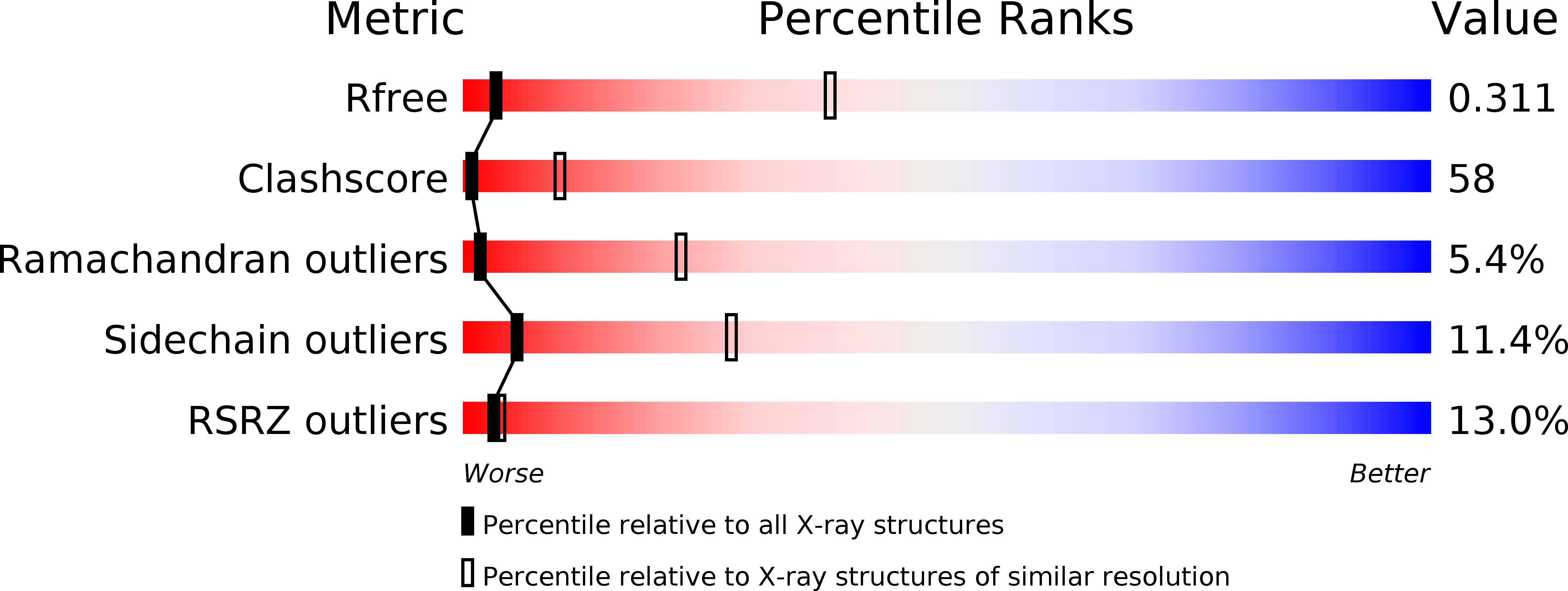
Resolution:
3.80 Å
R-Value Free:
0.33
R-Value Work:
0.27
R-Value Observed:
0.28
Space Group:
I 2 2 2
Macromolecular Entities
Polymer Type:polypeptide(L)
Description:FAB
Chain IDs:A, C
Chain Length:215
Number of Molecules:2
Biological Source:Mus musculus
Polymer Type:polypeptide(L)
Description:FAB
Chain IDs:B, D
Chain Length:224
Number of Molecules:2
Biological Source:Mus musculus
Polymer Type:polypeptide(L)
Description:Voltage-gated potassium channel
Chain IDs:E (auth: K), F (auth: L), G (auth: M), H (auth: N)
Chain Length:139
Number of Molecules:4
Biological Source:Streptomyces lividans
Ligand Molecules
Primary Citation
Crystal structure of full-length KcsA in its closed conformation.
Proc.Natl.Acad.Sci.USA 106 6644 6649 (2009)
PMID: 19346472 DOI: 10.1073/pnas.0810663106

Abstact

KcsA is a proton-activated, voltage-modulated K(+) channel that has served as the archetype pore domain in the Kv channel superfamily. Here, we have used synthetic antigen-binding fragments (Fabs) as crystallographic chaperones to determine the structure of full-length KcsA at 3.8 A, as well as that of its isolated C-terminal domain at 2.6 A. The structure of the full-length KcsA-Fab complex reveals a well-defined, 4-helix bundle that projects approximately 70 A toward the cytoplasm. This bundle promotes a approximately 15 degree bending in the inner bundle gate, tightening its diameter and shifting the narrowest point 2 turns of helix below. Functional analysis of the full-length KcsA-Fab complex suggests that the C-terminal bundle remains whole during gating. We suggest that this structure likely represents the physiologically relevant closed conformation of KcsA.

Legend

Protein

Chemical

Disease

Primary Citation of related structures