3EF4 image
Deposition Date 2008-09-08
Release Date 2008-12-30
Last Version Date 2023-11-01
Entry Detail
PDB ID:
3EF4
Title:
Crystal structure of native pseudoazurin from Hyphomicrobium denitrificans
Biological Source:
Source Organism:
Method Details:
Experimental Method:
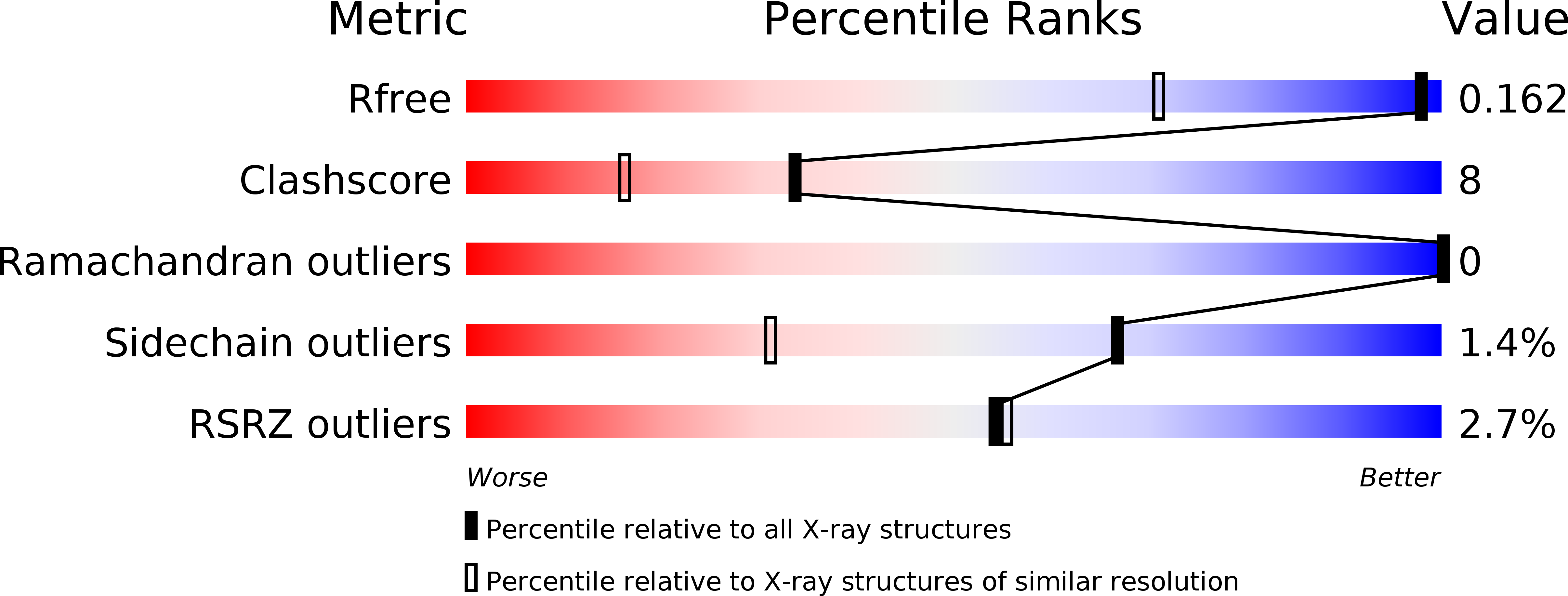
Resolution:
1.18 Å
R-Value Free:
0.16
R-Value Work:
0.13
R-Value Observed:
0.13
Space Group:
C 1 2 1
Macromolecular Entities
Structures with similar UniProt ID
Protein Blast
Polymer Type:polypeptide(L)
Molecule:Blue copper protein
Gene (Uniprot):paz
Chain IDs:A, B, C
Chain Length:124
Number of Molecules:3
Biological Source:Hyphomicrobium denitrificans
Primary Citation
Atomic resolution structure of pseudoazurin from the methylotrophic denitrifying bacterium Hyphomicrobium denitrificans: structural insights into its spectroscopic properties
Acta Crystallogr.,Sect.D 65 85 92 (2009)
PMID: 19153470 DOI: 10.1107/S0907444908040195

Abstact

The crystal structure of native pseudoazurin (HdPAz) from the methylotrophic denitrifying bacterium Hyphomicrobium denitrificans has been determined at a resolution of 1.18 A. After refinement with SHELX employing anisotropic displacement parameters and riding H atoms, R(work) and R(free) were 0.135 and 0.169, respectively. Visualization of the anisotropic displacement parameters as thermal ellipsoids provided insight into the atomic motion within the perturbed type 1 Cu site. The asymmetric unit includes three HdPAz molecules which are tightly packed by head-to-head cupredoxin dimer formation. The shape of the Cu-atom ellipsoid implies significant vibrational motion diagonal to the equatorial xy plane defined by the three ligands (two His and one Cys). The geometric parameters of the type 1 Cu site in the HdPAz structure differ unambiguously from those of other pseudoazurins. It is demonstrated that their structural aspects are consistent with the unique visible absorption spectrum.

Legend

Protein

Chemical

Disease

Primary Citation of related structures
Feedback Form
Name
Email
Institute
Feedback