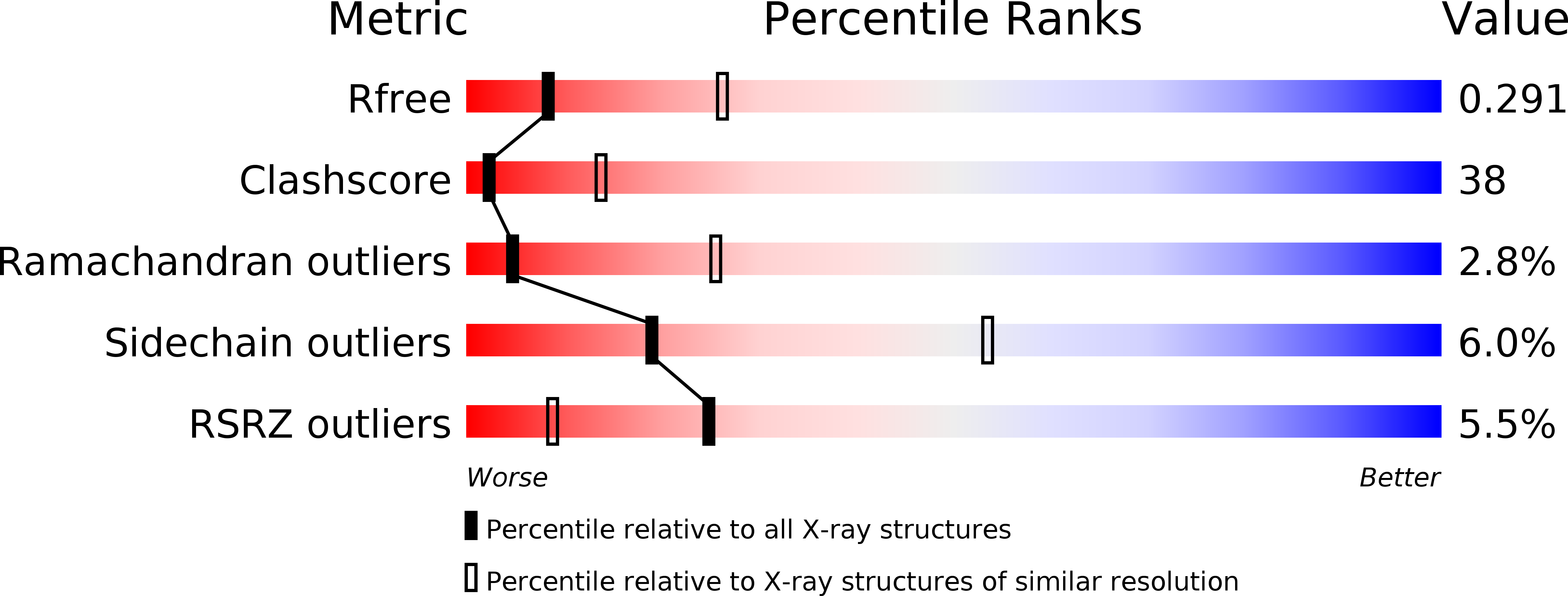
3EEC image
Deposition Date 2008-09-04
Release Date 2009-03-10
Last Version Date 2023-11-01
Entry Detail
PDB ID:
3EEC
Title:
X-ray structure of human ubiquitin Cd(II) adduct
Biological Source:
Source Organism:
Homo sapiens (Taxon ID: 9606)
Host Organism:
Method Details:
Experimental Method:
Resolution:
3.00 Å
R-Value Free:
0.30
R-Value Work:
0.27
R-Value Observed:
0.27
Space Group:
P 43 3 2
Macromolecular Entities
Polymer Type:polypeptide(L)
Molecule:Ubiquitin
Chain IDs:A, B
Chain Length:76
Number of Molecules:2
Biological Source:Homo sapiens
Ligand Molecules
Primary Citation
Structural probing of Zn(II), Cd(II) and Hg(II) binding to human ubiquitin.
Chem.Commun.(Camb.) 45 5960 5962 (2008)
PMID: 19030552 DOI: 10.1039/b813463d

Abstact

A structural investigation performed on adducts of human ubiquitin with group-12 metal ions reveals common preferential anchoring sites, the most populated one being His68; at higher metal ion concentration a second and a third site, close to the N-terminus of the protein, become populated and promote a polymorphic transition from orthorhombic to cubic form; Glu16 and Glu18, involved in the latter metal binding, undergo a remarkable displacement from their position in native ubiquitin; the aggregate stereochemistry appears to be driven by the clustering of deshielded backbone hydrogen-bond patches, and metal ions foster this process.

Legend

Protein

Chemical

Disease

Primary Citation of related structures