Deposition Date
2008-09-03
Release Date
2008-11-18
Last Version Date
2024-10-30
Entry Detail
PDB ID:
3EDY
Keywords:
Title:
Crystal Structure of the Precursor Form of Human Tripeptidyl-Peptidase 1
Biological Source:
Source Organism(s):
Homo sapiens (Taxon ID: 9606)
Expression System(s):
Method Details:
Experimental Method:
Resolution:
1.85 Å
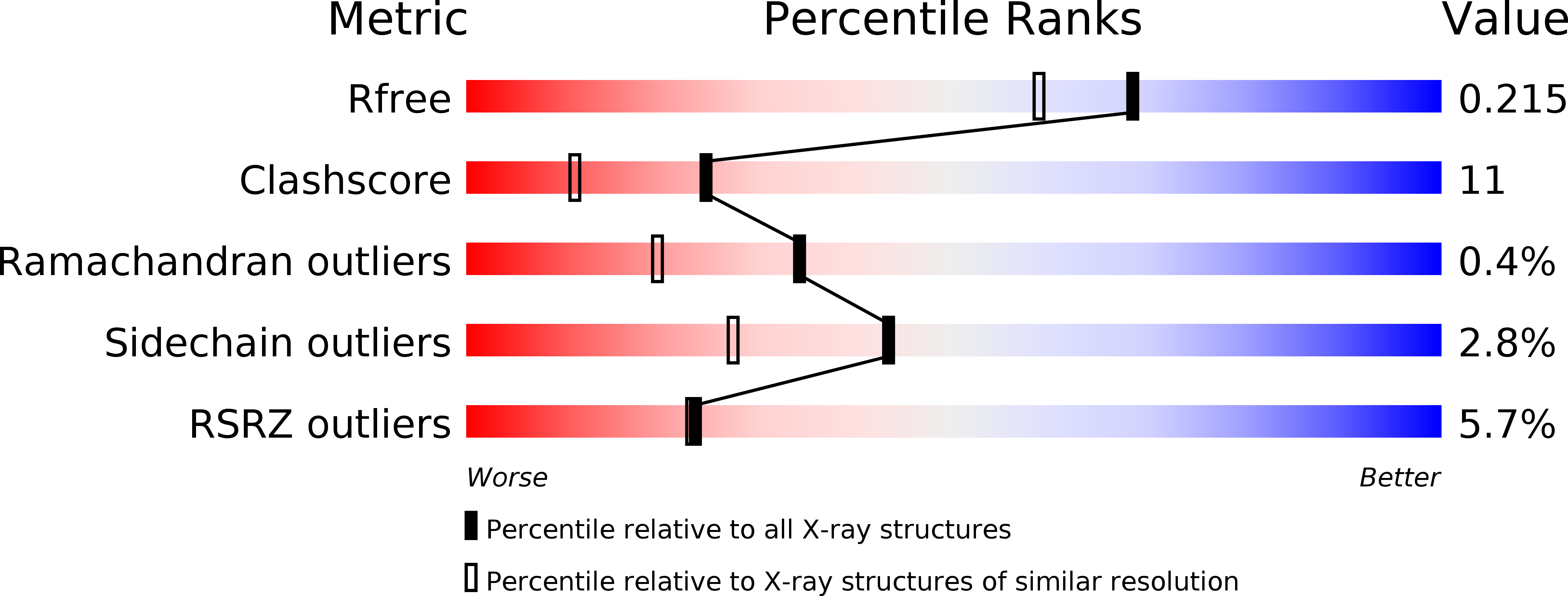
R-Value Free:
0.20
R-Value Work:
0.17
R-Value Observed:
0.17
Space Group:
P 21 21 21