Deposition Date
2008-09-02
Release Date
2008-11-04
Last Version Date
2024-11-20
Entry Detail
PDB ID:
3ED3
Keywords:
Title:
Crystal Structure of the Yeast Dithiol/Disulfide Oxidoreductase Mpd1p
Biological Source:
Source Organism(s):
Saccharomyces cerevisiae (Taxon ID: 4932)
Expression System(s):
Method Details:
Experimental Method:
Resolution:
2.00 Å
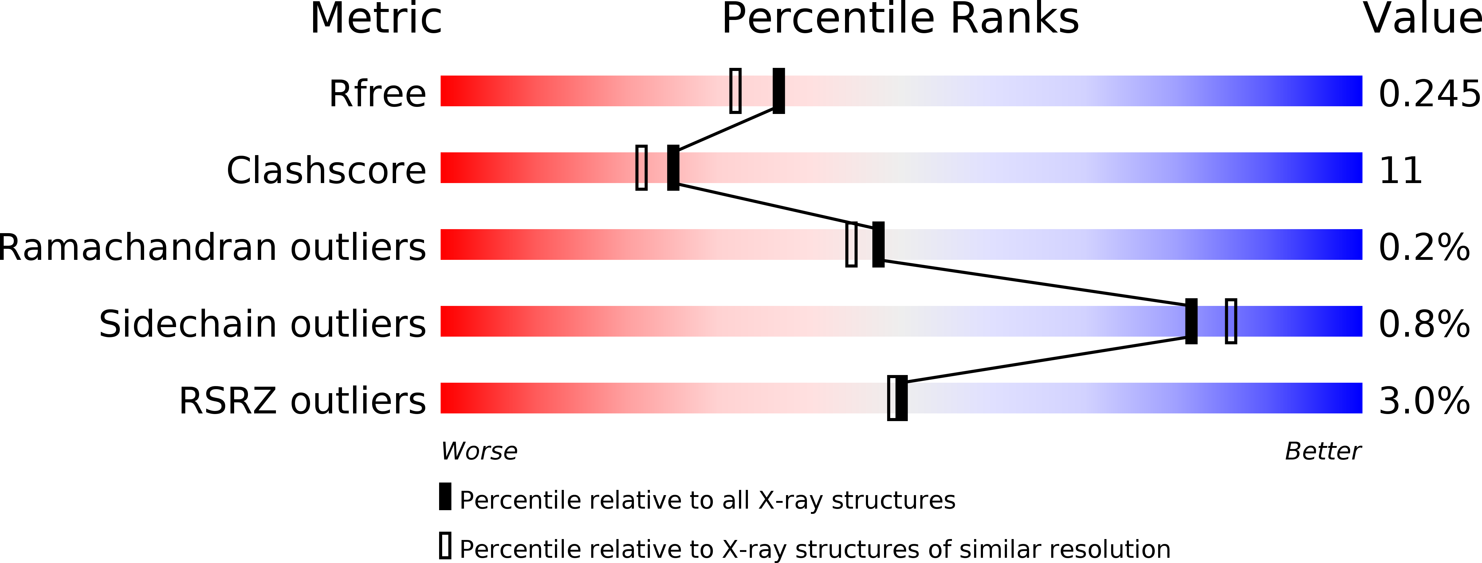
R-Value Free:
0.25
R-Value Work:
0.21
Space Group:
P 1 21 1