Deposition Date
2008-08-27
Release Date
2008-12-09
Last Version Date
2024-02-21
Entry Detail
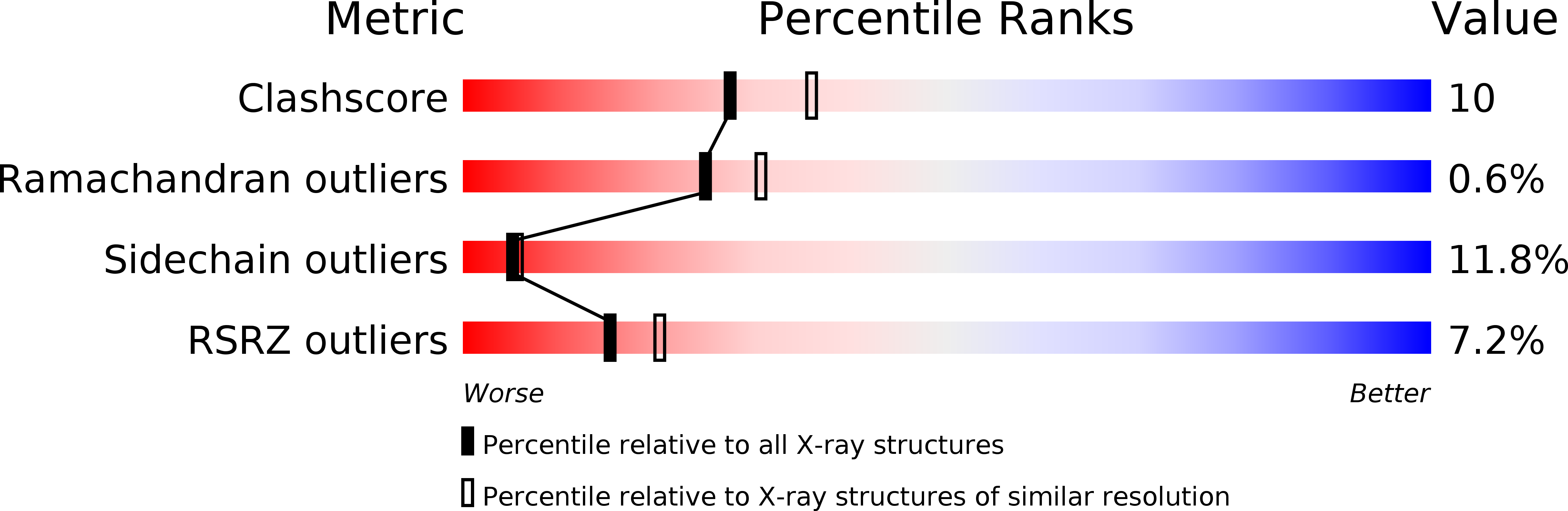
PDB ID:
3EBE
Keywords:
Title:
Crystal structure of xenopus laevis replication initiation factor MCM10 internal domain
Biological Source:
Source Organism(s):
Xenopus laevis (Taxon ID: 8355)
Expression System(s):
Method Details:
Experimental Method:
Resolution:
2.30 Å
R-Value Free:
0.24
R-Value Work:
0.20
R-Value Observed:
0.20
Space Group:
P 1 21 1