3E9J image
Deposition Date 2008-08-22
Release Date 2008-11-25
Last Version Date 2024-10-30
Entry Detail
PDB ID:
3E9J
Keywords:
Title:
Structure of the charge-transfer intermediate of the transmembrane redox catalyst DsbB
Biological Source:
Source Organism:
Host Organism:
Method Details:
Experimental Method:
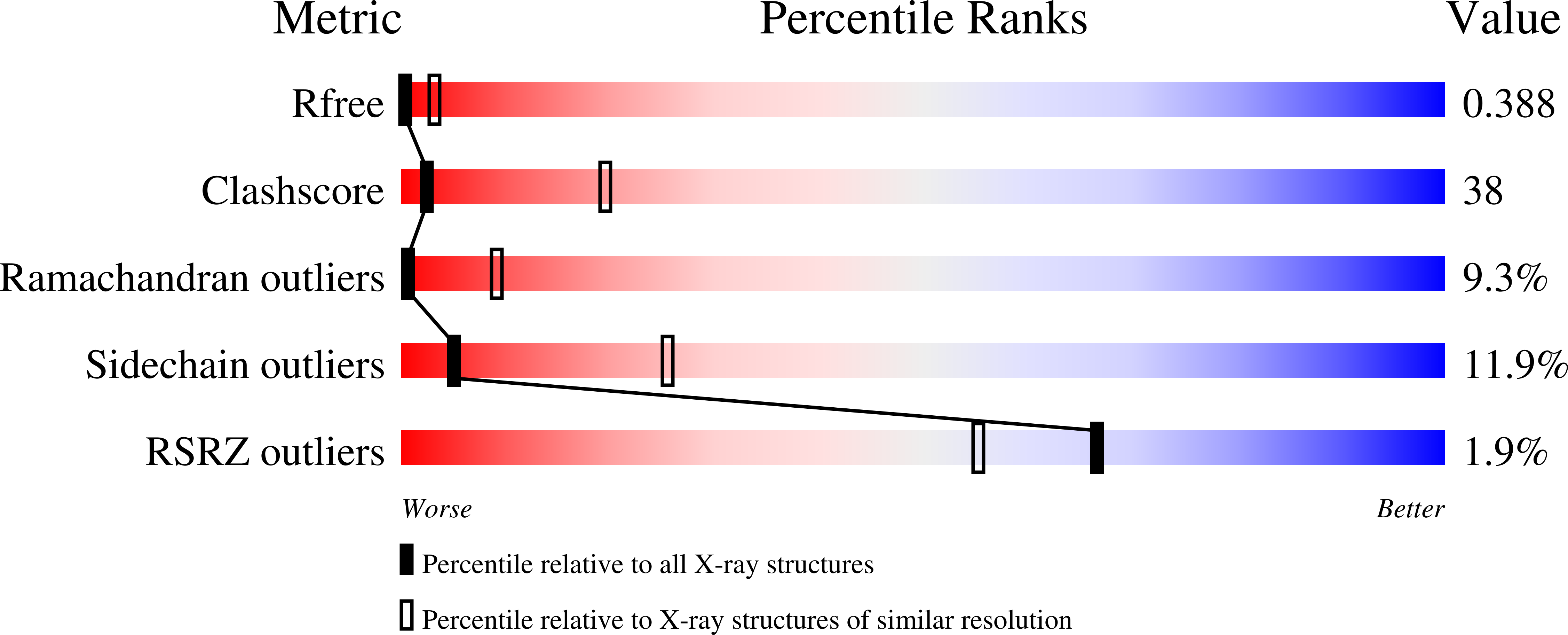
Resolution:
3.70 Å
R-Value Free:
0.37
R-Value Work:
0.33
R-Value Observed:
0.33
Space Group:
P 1 21 1
Macromolecular Entities
Structures with similar UniProt ID
Protein Blast
Polymer Type:polypeptide(L)
Molecule:Thiol/disulfide oxidoreductase DsbA
Gene (Uniprot):dsbA
Mutagens:C33A
Chain IDs:A (auth: B), C (auth: E)
Chain Length:189
Number of Molecules:2
Biological Source:Escherichia coli K12
Structures with similar UniProt ID
Protein Blast
Polymer Type:polypeptide(L)
Molecule:Thiol/disulfide oxidoreductase DsbB
Gene (Uniprot):dsbB
Mutagens:C8A, C49V
Chain IDs:B (auth: C), D (auth: F)
Chain Length:182
Number of Molecules:2
Biological Source:Escherichia coli K12
Ligand Molecules
Primary Citation
Preparation and structure of the charge-transfer intermediate of the transmembrane redox catalyst DsbB.
Febs Lett. 582 3301 3307 (2008)
PMID: 18775700 DOI: 10.1016/j.febslet.2008.07.063

Abstact

Disulfide bond formation is a critical step in the folding of many secretory proteins. In bacteria, disulfide bonds are introduced by the periplasmic dithiol oxidase DsbA, which transfers its catalytic disulfide bond to folding polypeptides. Reduced DsbA is reoxidized by ubiquinone Q8, catalyzed by inner membrane quinone reductase DsbB. Here, we report the preparation of a kinetically stable ternary complex between wild-type DsbB, containing all essential cysteines, Q8 and DsbA covalently bound to DsbB. The crystal structure of this trapped DsbB reaction intermediate exhibits a charge-transfer interaction between Q8 and the Cys44 in the DsbB reaction center providing experimental evidence for the mechanism of de novo disulfide bond generation in DsbB.

Legend

Protein

Chemical

Disease

Primary Citation of related structures